Microscopy Imaging Techniques
Microscopy imaging techniques are employed by scientists and researcher to improve their ability to view the microscopic world.
Advances in microscopy enable visualization of a broad range of biological processes and features in cell structure. This page outlines the different types and provides a brief introduction to the technique.
You are invited to follow the corresponding links to read further about the technique you are interested in reviewing.

Brightfield Microscopy - is the most elementary form of imaging a specimen and is generally used with compound microscopes. The technique takes the specimen which is dark and contrasts it by the surrounding bright viewing field. Compound light microscopes are often simply referred to as brightfield microscopes. Right:algae with visible cells
Oil Immersion Microscopy - Oil Immersion Microscopy is an essential tool in examining specimens under a compound microscope. Although its few disadvantages are significant, careful technique will minimize problems such as cement drying on a lens. Similar refractive indexes allow for large bright images, especially useful to the study of inanimate objects, striated tissue and bacteria; a mixture of synthetic oils can create the most suitable viscosity to achieve high resolute quality images.
Kohler Illumination - is a microscopy imaging technique first developed in 1893 through the optimization of a microscope’s optical train so as to allow for homogenously bright light without artifacts and glare.

Darkfield Microscope - is the optimum microscopy technique for making objects appear bright against a dark background otherwise their refractive values are similar to the background and they will not be properly imaged. Dark Field is achieved by modifying your microscope. Right:sugar crystals
Differential Interference Contrast microscopy - is a microscopy imaging technique which benefits from differences in the light refraction by different sections of living cells and transparent specimens and allows for better visibility during microscopic evaluation.

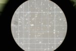
Phase Contrast Microscope - is most useful in viewing “phase objects” which are transparent, colorless and/or unstained specimens. This is employing a microscopy imaging technique benefiting molecular and cellular biology, microbiology and medical research. You are invited to explore the applications, pros and cons of this technique. At right: hemocytometer with fibroblasts.
See Phase Contrast Microscopy images
Fluorescence Microscope - employs high-powered light waves to provide unique image viewing options that are unavailable with conventional light microscopes. This imaging technique also uses stains so as to better view more components and details of the inner structures of cells. See immunofluorescence technique also.

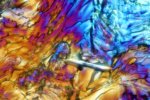
Polarizing Microscope - the ideal choice for birefringent materials. Polarization is used to enhance contrast and color to images providing information about absorption, structure and composition of specimens. The study of rocks and minerals in geology or petrography fields benefits through the use of this imaging technique as well as medicine, biology and metallurgy. At right: microscopic artificial sweetener crystals in polarized light.

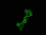
Confocal Microscope - Separating light waves with lasers and with the use of state of the art technology, images can be viewed without blurred edges and in higher resolutions. Many images can be taken quickly with a small section of the sample viewed at a time. At right: immunohistochemical stain of vimentin protein in smooth muscle cells.
See Super-Resolution Microscopy
Enjoy!!
Return from Microscopy Imaging Techniques to Best Microscope Home
Find out how to advertise on MicroscopeMaster!


