Yeast Cells Under the Microscope
** Characteristics, Habitat and Observation
What is Yeast?
Yeast cells are members of the Fungus Kingdom. They are single celled microorganisms (eukaryotic) classified under phyla Ascomycota (sac fungi) and Basidiomyota (higher fungi) both of which fall under the subkingdom Dikarya.
Yeast Cells
- Kingdom - Fungus
- Subkingdom - Kikarya
- Phyla - Ascomycota and Basidiomycota
* Budding yeast, also referred to as true yeast fall under the Phylum Acomycota and in the Order Saccharomycetales
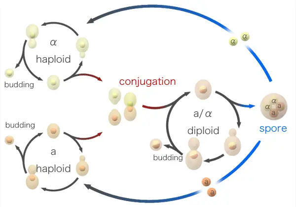
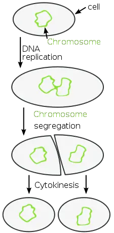
Yeast are very diverse (over 1,500 species) with most forming the phylum Ascomycota while only a few are classified as Basidiomycota. Yeast cells reproduce through budding or binary fission which are both methods of asexual reproduction (Horst, 2010).
Budding - A new yeast cell is formed through mitotic cell division and remains attached as a bud on the old cell until it splits and becomes independent. Here, the parent cell produces an outgrowth that finally splits to become an independent identical cell as the parent cell.
Binary Fission - In binary fission, no outgrowth (bud) is formed. Rather, through mitosis, the genome replicates and divides followed by the formation of a new plasma membrane and ultimately the cell dividing in to two to form two new cells from the parent cell.
* Some of the fungi referred to as dimorphic tend to alternate between the yeast and hyphal phase which means that they can also grow as hyphae (thread like)
Habitat
Because they are very diverse, yeast can be found in a wide variety of habitats particularly in environments with sugar-rich materials. They are likely to be found on flowers, plant leaves, and fruits as well as on soil, deep-sea environments, skin surface and even intestinal tracts of animals (warm blooded).
While they can be found in many
environments, yeast require moist environments with sufficient amounts of simple
and soluble nutrients to support growth and multiplication (Horst, 2010).
While such yeast as the Candica can cause infections (Candidiasis) there are useful yeast such as:
- Baker’s Yeast
- Nutritional Yeast
- Brewer’s Yeast
- Distiller’s and Wine Yeast
Yeast Under the Microscope
Requirements
- Water
- Yeast cake
- Dropper
- Glass slides and cover slips
- Stains (stated below)
Yeast cake contains Saccharomyces Cerevisiae (sugar-eating fungus) and can therefore be used to obtain the yeast to observe under the microscope. The following is a procedure that can be used to prepare the specimen for observation.
- Obtain yeast cake (this can be bought from bakery specialty stores or a supermarket)
- Cut a small piece of yeast cake and mix with water to form a pasty texture
- Add a little more water to form a solution
- Using a dropper, collect and place a drop of the solution on a microscope slide
- Place a microscope cover slip and observe under high power objective
Observation
With Brightfield Microscopy
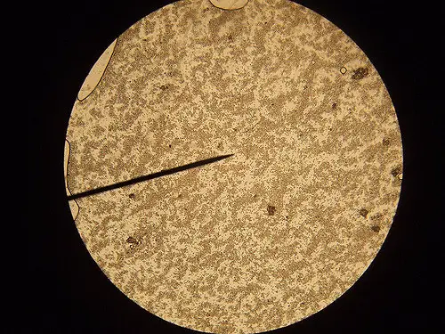
When viewing the specimen under high
magnification (1000x and above) one will see oval (egg shaped) organism, which
are the yeast. It is also possible to observe the buds, which can be seen on
some of the yeast cells.
If the solution had some sugar, one will also notice some bubbles in the specimen, which are as a result of the fermentation process by the microorganisms.
With Fluorescence Microscopy
Fluorescence microscopy can be used for the purposes of observing the organelles inside the yeast cells. This is particularly a great method through which students can get to view the intracellular distribution of the cell and identify the different types of cell organelle.
This may prove a little challenging when it comes to yeast given that they are very small in size (compared to other cells) and have a cell wall (Chalfie and Kain, 2005).
For living yeast cells, a number of dyes have to be used in order to increase contrast and be able to differentiate the different organelles in the cells.
These include:
- DAPI for the nuclei
- FM4-64 for the vacuoles
- DIOC6 for the endoplasmic reticulum and mitochondria
- DASPMI for mitochondria
- Calcofluor White which helps stain the cell wall
Yeast are among the smallest eukaryotic cells with diameters of between 5 and 10um. For this reason, it is important to view them under high magnification using fluorescence microscopy.
Here, 60x or 100x objectives with numerical aperture of 1.4 are recommended for visual observation and maximum brightness and resolution respectively.
If recording using a digital camera (e.g. using a 6.8 x 6.8 um sq pixel camera) then a magnification of 60x would be recommended with numerical aperture of 1.4 (Hašek, 2006).
See also:
Mycology in general as field of study
Fungi - Types, Morphology and Structure, Uses and Disadvantages
Taking a further look at Mold under the Microscope.
Return from Yeast Under the Microscope to Microscope Experiments
Return to MicroscopeMaster Home
Sources
Martin Chalfie and Steven R. Kain (2005) Green Fluorescent Protein: Properties, Applications and Protocols.
Jiří Hašek (2006) Yeast Fluorescence Microscopy. Volume 313 of the series Methods in Molecular Biology pp 85-96.
Feldmann, Horst (2010). Yeast. Molecular and Cell bio.
Find out how to advertise on MicroscopeMaster!


