Endoscopes
Parts, Uses and New Technologies
What is an Endoscope?
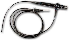
Essentially, an endoscope may be described as a long, thin illuminated flexible tube that has a camera on one end. Today, the endoscope has become of the most important devices in medicine serving to view the inside of body cavities.
Although endoscopes are usually inserted through such openings as the mouth and the rectum, they are also inserted into the body through small incisions on the skin particularly during keyhole surgery (minimal invasive surgery).
Parts of an Endoscope
A standard endoscope in composed of the following parts:
- A thin, long flexible tube
- A lens or lens system
- A light transmitting system
- The eyepiece
- Control system
How the Endoscope Works
Basically, a typical endoscope uses fiber optics, which allow for effective transmitting of light. In this technique (fiber optics) light is transmitted through a flexible fiber of glass (transparent) known as optical fiber(s).
The optical fiber allows for light to travel through curved paths, which makes one of the best systems to view spaces that would normally be difficult to reach. Here, total internal reflection makes it possible for light to travel along the fibers with the light rays hitting the fiber walls at an angle (minimum angle of 82 degree).
Given that individual fibers can be thinner than human hair, fiber optics is one of the best techniques to enter and view different areas of the body.
There are typically two sets of the fibers. These include the outer fiber that functions to supply light and an inner coherent ring that serves to transmit the image.
The outer fiber - This fiber contains a number of fibers that have been bundled together in no particular order. It is for this reason that the outer fiber is commonly referred to as the incoherent bundle. The fiber is entirely enclosed with a sleeve to protect it. Typically, it is coated with either plastic or steel which protects it from water or moisture (making it waterproof).
Inner fiber - Like the outer fiber, the inner bundle is also composed of a bundle of fibers. However, unlike the outer bundle, the inner fiber is in perfect order, which is why it is referred to as the coherent bundle. The tiny lens connected to the end of this bundle allows for light to be effectively focused so that reflected light from the object of interest can be collected and transmitted for viewing.
Other Important Parts
- Water pipes - The pipes serve to carry water which is used to wash the lens thereby maintaining a clear view.
- The operational channel - This is an opening on the device that is used to move various accessories to the distil end (of the endoscope) for surgery purposes.
- Control cables - This is used to control the direction that the distil end will bend as it moves through body cavities.
New Endoscope Technologies:
Wireless Capsule Endoscopy
Capsule endoscopy is one of the new procedures
that involve the use of a very small wireless camera to take pictures in the
digestive system.
For this procedure, one swallows a capsule the size of vitamin-sided capsule or a large pill. The technology involves the use of a wireless miniature encapsulated camera that takes pictures as the capsule travels through the digestive system.
As it travels down the digestive system, the capsule wirelessly transmit the images it captures which can then be used to detect any issues in the digestive tract. The images (it can take thousands of images) are then transmitted to a recorder from which they can be retrieved. Like ingested food, the capsule travels through the digestive system and ultimately leaves the body when the individual passes stool.
The main components of capsule endoscopy include:
- Sensor array (electrodes) - The patient wears this around the abdomen area like a sensor belt
- Data recorder worn by the patient and connected to the electrodes
- The capsule that is 26mm by 11mm in size - Some of the components of the capsule include; a lens, diodes (that emit light) a semi-conductor, an antenna as well as a transmitter.
Since its approval by the FDA in 2001, capsule endoscopy has been shown to be an effective procedure with a number of advantages that include:
- painless
- disposable
- non-invasive
Confocal Laser Endomicroscopy and Endocytoscopy
These are some of the new procedures aimed at enhanced high resolution in the assessment of gastrointestinal mucosal histology at both the cellular and sub-cellular level.
Basically, the technique is based on the principle of illuminating the tissue of interest with low power laser which in turn allows for the detection of fluorescent light that is reflected from the tissue.
With this procedure, it becomes possible to carry
out in vivo examinations with images being displayed in real-time. It has been
shown to be particularly beneficial in the detection of abnormal growth of
tissue in conditions like ulcerative colitis.
Uses of Endoscopes
Although endoscopy is largely used for the purposes of examining a patient's digestive tract, endoscopes are also used for:
Arthroscopy - This is a medical, surgical procedure that is used to visualize joints, identify the problem and start treatment. For this procedure, the surgeon typically makes a small incision into the skin of the patient and inserts the arthoscope to visualize the joint. The procedure is particularly useful in the diagnosis of injuries to the joint as well as any diseases that may affect the joints.
Bronchoscopy - In bronchoscopy, the healthcare professional uses a bronchoscope to visualize the airway. The procedure allows the doctor to closely examine all the parts of the airway including the throat, the larynx as well as the trachea. Bronchoschopy is divided into flexible and rigid bronchoscopy.
Whereas a long, thin and flexible tube is used in flexible bronchoscopy, a straight and hollow metal tube is used in rigid bronchoscopy and requires the use of general anesthesia unlike with flexible bronchoscopy. Bronchoscopy is used to detect any problems with the airway system and thus treat the problem.
Endoscope Biopsy - Using this procedure, the physician inserts an endoscope through a body opening or a tiny incision to reach the area of interest. By using biopsy forceps, the physician/surgeon can then obtain a tissue sample from the body area for analysis.
Laparoscopy - This is a procedure where the endoscope is inserted into the body through a tiny incision for the visualization of various abdominal organs as well as surgery if need be.
Return to Surgical Microscopes
Return from learning about Endoscopes to MicroscopeMaster Home
Find out how to advertise on MicroscopeMaster!

![Endoscope Training by Georg Graf von Westphalen [CC BY 3.0 (http://creativecommons.org/licenses/by/3.0)], via Wikimedia Commons Endoscope Training by Georg Graf von Westphalen [CC BY 3.0 (http://creativecommons.org/licenses/by/3.0)], via Wikimedia Commons](https://www.microscopemaster.com/images/Endoscopetraining.png)


