Virology
Overview, Classification, Diseases - Clinical, Veterinary
Definition: What is Virology?
Essentially, virology is the branch of microbiology that deals with the study of viruses (as well as various virus-like particles), their characteristics, classification, as well as the relationship with their respective hosts.
Compared to the other organisms in microbiology, viruses are very unique with different characteristics (with regards to multiplication, structure, etc) that set them apart.
Given that viruses are of medical and veterinary significance, virology has increasingly become one of the most important sub-disciplines of microbiology that has allowed researchers to not only discover treatments and cures for the diseases they cause, but also use them for pharmaceutical purposes.
Some of the general characteristics of a virus include:
- Can only reproduce (through synthesis and assembly) in living cells
- Contain DNA/RNA or both in some cases
- Are not capable of sexual or asexual modes of reproduction
- Are not cells - They are acellular particles that lack normal cell organelles and cytoplasm
- Very small compared to other single-celled organisms
* Unlike other true single-cells organisms, viruses are referred to as "particles" in most books because they are not considered to be "living" cells.
* They are parasites that fully depend on living cells for replication.
Virus Classification
According to a system that was proposed in the 70s by the International Committee on the Taxonomy of Viruses (ICTV), the classification (naming) was provided:
- Phylum: Viricota
- Class: Viricetes
- Order: Virales
- Family: Viridae
- Subfamily: Virinae
- Genus: Virus
- Subgenus: Virus
Apart from this system of classification, virology also classifies viruses based on the following characteristics:
Nature of Nucleic Acid
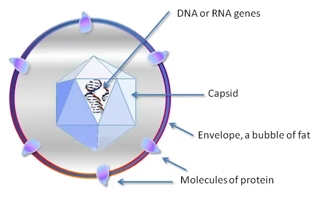
For the most part, cells of living organisms contain DNA in their nucleus that carries the genetic material. For viruses, however, they either carry DNA or RNA with genes that are responsible for encoding specific proteins.
The DNA and RNA between various types of viruses are also different which allows specific viruses to be identified. Whereas Poxviruses and Herpes viruses contain an enveloped, double-stranded DNA, the double-stranded DNA of Adenoviruses and Polyomaviruses do not have an envelope while Parvoviruses contain unenveloped, single-stranded DNA.
These differences are also observed in RNA viruses. For instance, such viruses as Retroviruses and Togaviruses contain enveloped RNA while Picomaviruses lack this outer envelope. As well, the RNA of such viruses as Reoviruses is contained in a double capsid.
In order to direct the synthesis of proteins, viral RNA first encodes enzymes that replicate the RNA to DNA. The new DNA molecule is then directly responsible for the synthesis of viral proteins.
Some of the other structures of the genetic material may take the following forms:
- Linear - E.g. smallpox and rabies viruses
- Circular - E.g. Papillomaviruses
- Non-segmented - E.g. Parainfluenza viruses
- Segmented - E.g. Influenza viruses
Symmetry of Protein Shell
Different types of viruses also have different shape/morphology. Currently, several shapes of the virus shell have been identified, which has in turn been used to classify different types of viruses.
These include:
· Helical symmetry - Viruses with this morphology contain a layer of capsomer that is stacked around the nucleic acid forming a helical shape. Assembly of the protein subunits forms an elongated helical structure that is either flexible or tough in nature. Examples of these viruses include the Sendal virus and the tobacco mosaic virus.
· Icosahedral Symmetry - Typically, viruses classified under this morphology have a polyhedron structure consisting of about 20 faces/sides of equilateral triangles as well as 12 verticles/corners. Here, lines that run through the opposite vertices define the general appearance of the shell.
For instance, those that run through the centers of the faces of opposite triangles result in threefold rotational symmetry axes. Here, then, icosahedra symmetry may range from fivefold to twofold rotational symmetry. Adenovirus, rhinovirus, and poliovirus are good examples of viruses that fall in this category.
· Prolate - This is a type of icosahedra that is elongated. As a result, they may appear more cylindrical in shape given that the elongation is along one axis. This morphology has been associated with a majority of viruses known as bacteriophages (virus that infects and replicate in bacteria). Good examples of bacteriophages include the M13 bacteriophage and Escherichia Virus T4.
· Complex - The complex structure is a combination of the helical and icosahedral symmetry. Therefore, viruses with capsids referred to as complex cannot be fully classified as helical or icosahedral.
In some cases, these viruses may contain additional structures such as a complex cell wall. This allows for the virus to be easily identified based on any extra features. Using such extra structures as the helical tail, the virus can attach on to a cell (e.g. bacteria) before inserting their DNA. The Poxvirus that causes smallpox in human beings is an example of a virus with a complex shell.
· Envelope - Compared to the other shells, the shell of viruses, referred to as an envelope, are covered by a lipid bilayer membrane. In most cases, this covering is formed as the virus exits the host cell. Some of the viruses that have the lipid bilayer envelope include the HIV and Influenza virus.
Lipid Membrane: Presence or Absence
For some viruses, the envelope has been shown to consist of a lipid bilayer. This envelope, however, comes from the cell lipids of the host and not the virus itself. Given that this structure is only present in some viruses, it is used for classification purposes.
For viruses with the lipid bilayer, the envelope serves a number of roles that support infection. For instance, some of the functions of the lipid bilayer include attachment, content release into the cell as well as the packaging of newly assembled particles.
Flaviviridae, HIV, H. influenzae and many other animal viruses are examples of particles that contain a lipid bilayer envelope. Some of the viruses that lack this covering (unenveloped) include Caliciviridae and Papillomaviridae.
Dimension of the Capsid or Virion
Viruses are also classified based on the dimensions of the capsid of the virion (the entire virus including the outer and inner shell). According to studies, the mean radii of a virus significantly vary (from about 10 to 200nm). This allows for different types of viruses to be classified solely based on these dimensions.
The Baltimore System
Apart from classification based on the aforementioned characteristics, viruses are also classified in groups based on the Baltimore classification system. Developed by David Baltimore, a Nobel Prize-Winning biologist, this is one of the most commonly used systems for virus classification.
This classification system groups viruses based on mRNA production:
· Group I - Includes viruses (E.g. the herpesvirus) with double-stranded DNA that produces mRNA through transcription. Here, the virus uses enzymes belonging to the host.
· Group II - Include those with single-stranded DNA (E.g. Parvovirus). This is first converted into the double-stranded intermediate before the mRNA is produced.
· Group III - These viruses (E.g. Rotavirus) have a double-stranded RNA. One of the strands acts as the template for mRNA generation. Here, the enzyme encoded by the virus is involved in mRNA generation.
· Group IV - This group includes viruses with a single-stranded RNA (E.g. Picornavirus). Although this RNA can serve as the mRNA, double-stranded RNA (replicate intermediates) are first produced to produce mRNA.
· Group V - Group V viruses (E.g. Rhabdovirus) contain single-stranded RNA. Unlike Group IV viruses, however, their RNA cannot directly act as mRNA and are therefore complementary. However, double-stranded RNA is first produced before the production of mRNA.
· Group VI - Viruses in this group (E.g. HIV virus) contain a diploid single-stranded RNA that is first converted to a double-stranded DNA before the production of mRNA.
· Group VII - Viruses of Group VII (E.g. Hepadnavirus) have a partially double-stranded DNA. These genomes first name single-stranded RNA intermediates that also act s mRNA.
Clinical Vs Veterinary Virology
Veterinary virology, which is a branch of virology concerned with the viral agents, animal viral diseases and any emerging zoonotic diseases caused by viruses, developed from a need to control viral diseases in animals (e.g. prion diseases, pestivirus, arterivirus, etc).
While veterinary virology is an important field of study that is aimed at preventing and treating animal diseases caused by viruses, it's also an important field in clinical virology. This is due to the fact that such diseases as rabies that affects canines can also affect human beings.
Clinical Virology, on the other hand is a field of medicine and virology that is concerned with the study of viruses that cause human pathologies. Like veterinary virology, clinical virology is also concerned with the classification and characterization of these particles, which has, in turn, made it possible to develop treatment and prevention strategies against the diseases they cause.
Check out these overviews:
Return to Viruses under the Microscope
Return from Virology to MicroscopeMaster home
References
Akira Ono. (2010). Viruses and Lipids. ncbi.
Hans R. Gelderblom. (1996). Structure and Classification of Viruses. http://gsbs.utmb.edu/microbook/ch041.htm.
John Carter, Venetia Saunders, Venetia A. (2013). Virology: Principles and Applications.
M. A. Oyekunle, O. E. Ojo, and M. Agbaje. Introduction to Veterinary Microbiology.
Ting Chen1 and Sharon C. Glotzer. (2007). Simulation studies of a phenomenological model for elongated virus capsid formation.
Links
https://msu.edu/course/mmg/569/Virus%20Structure.htm
https://opentextbc.ca/biology2eopenstax/chapter/viral-evolution-morphology-and-classification/
Find out how to advertise on MicroscopeMaster!


![Helical, icosahedral and complex structures of viruses by CNX OpenStax [CC BY 4.0 (https://creativecommons.org/licenses/by/4.0)] Helical, icosahedral and complex structures of viruses by CNX OpenStax [CC BY 4.0 (https://creativecommons.org/licenses/by/4.0)]](https://www.microscopemaster.com/images/OSC_Microbio_06_01_virusshapes.jpg)


