What are Adipocytes?
What are their Functions?
Definition
Characterized by copious amounts of cytoplasmic lipid droplets, adipocytes are the primary components of adipose tissue that acts as energy reserves. While this is their primary function, they are also involved in a number of other important functions including the secretion of lipids as well as various protein factors.
Generally, these cells are divided into two main classes which include the white and brown adipocytes. However, there is a third type known as beige adipocytes. The different types originate from various progenitors and have several functions and characteristics.
Adipogenesis
Adipogenesis refers to the process through which adipocytes are produced from the differentiation of mesenchymal stem cells located in the bone marrow. This process starts with the production of preadipocytes following the commitment of the stem cells during the commitment phase.
Here, the stem cells, after they are activated, commit to the adipocyte lineage which means that ultimately, they can only give rise to adipocytes.
Following commitment of the mesenchymal stem cells; studies have revealed that there are multiple lineages that ultimately give rise to different types of adipocytes.
In the case of white adipocytes, preadipocytes (which are adipocyte progenitors) can be found in endothelial tissue, bone marrow, cartilage, as well as adipose tissue -Differentiation of these cells allow for white adipocytes only.
Brown adipocytes, on the other hand, have been shown to originate from the myogenic lineage. This means that their progenitors can be found in the cells of the muscle tissue.
Apart from this lineage, some studies have also shown that some progenitors of the white adipocytes can differentiate to become brown adipocytes. Here, stimulation of these preadipocytes resulted in adipogenesis to brown adipocytes.
As mentioned, stem cells have to be stimulated in order to commit and produce progenitors that in turn differentiate to give rise to the different types of adipocytes. This means that the process is regulated and thus requires signaling to occur.
Some of the signaling pathways involved in adipogenesis include:
- IGF-1 signaling (Insulin-Like Growth Factor 1)
- cAMP Signalling (Cyclic adenosine monophosphate signaling)
- BMP Signalling (Bone Morphogenetic Proteins signaling pathway)
- Wnt Signalling Pathway
- Hedgehog Signalling Pathway
- Glucocorticoid Signalling
- TGF-β Signalling (Transforming growth factor-β signaling)
Characteristics of Adipocytes
White Adipocytes
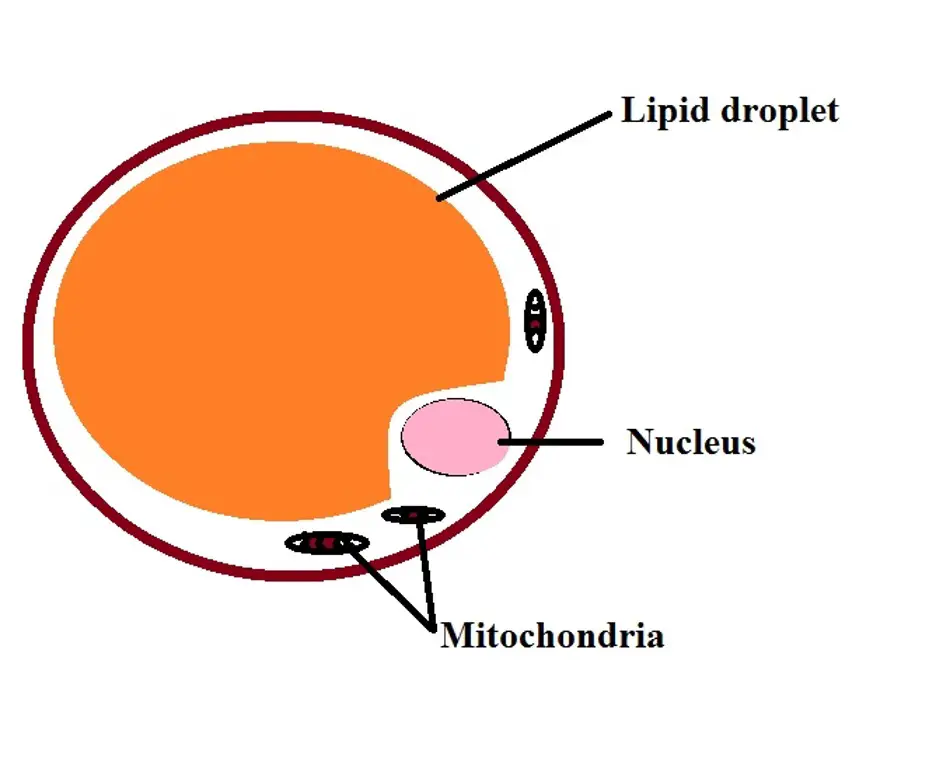
White adipocytes are characterized by a rounded (spherical) morphology and make up the white adipose tissue. Depending on their location, they may range from 25 to over 100um in diameter. Here, the general size of these cells is determined by the size of the lipid droplet in the cells.
While they contain a single droplet of lipid (therefore, referred to as univacuolar or unilocular adipocytes), it's large in size and can take more than 90 percent of the total cell volume. So then, the nucleus is pushed to one side of the cell. Due to the size of the droplet, white cells contain a small amount of cytoplasm with low-density of mitochondria.
As compared to brown adipocytes, white adipocytes are more abundant. As components of the white adipose tissue, white adipocytes are divided into two main groups depending on their distribution in the body.
Whereas visceral adipose consists of adipocytes that surround various body organs, subcutaneous adipose consists of adipocytes located under the skin.
Brown Adipocytes
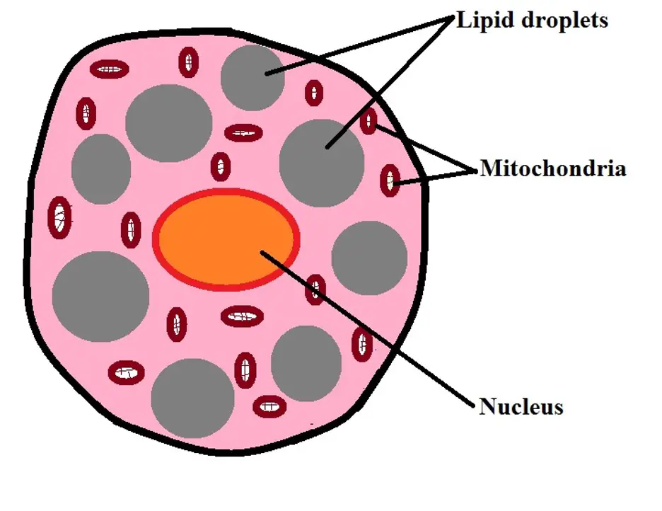
Unlike white adipocytes, brown adipocytes can only be found in mammals. They are characterized by a polygonal shape and are generally smaller in size, ranging from 15 to 60um in diameter. While they also contain lipid, like white adipocytes, brown adipocytes have multiple, vacuole-sized lipid droplets.
Given that the droplets do not occupy the central region of the cell, like in white adipocytes, the nucleus of brown adipocytes is centralized and not pushed to one side/pole of the cell. In addition to a centralized nucleus, brown adipocytes also have a larger volume of cytoplasm and numerous mitochondria when compared to the white ones. Because of the numerous mitochondria, these adipocytes appear darker in color.
Although brown adipocytes are commonly found in mammals, in human beings, studies have shown them to be absent in adults. However, they can be found in fetuses and newly born babies.
Located in the upper trunk, this group of cells is surrounded by a number of cells and tissues including the blood vessels, connective tissue as well as neurons of the sympathetic nervous system.
Beige Adipocytes
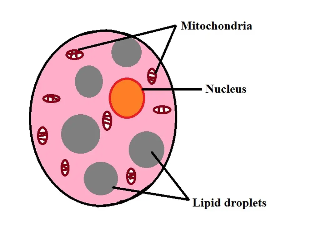
Beige adipocytes are similar to brown adipocytes in that they have multiple lipid droplets as such, they are also multilocular. For the most part, beige adipocytes (also known as brite adipocytes) are produced when white adipocytes transform and develop characteristics associated with the brown adipocytes.
This often occurs in response to various stimuli including exposure to cold, certain hormones (e.g. thyroid hormone, and some peptides etc). For this reason, beige adipocytes are considered members of the adaptive response.
* Beige adipocytes are characterized by a medium amount of mitochondria as well as relatively larger lipid droplets compared to those of brown adipocytes.
Function of White Adipocytes
As the most abundant adipocytes in the body, white adipocytes play a very important role in energy homeostasis. This is achieved through the accumulation of lipids that can then be broken down to produce energy when the body requires more.
For many multicellular organisms, consuming organic matter from the environment provides the energy required for survival. For these organisms, storing extra energy (in the form of lipids) in specialized cells is also important because it allows them to survive for a given period of time on reserves during periods of scarcity.
One of the biggest advantages of storing nutrients (source of energy) in the form of lipids is that generally lipids have higher calories. By breaking down lipids, more energy is produced than the energy that would be produced by breaking down other nutrients (e.g. carbohydrates).
Essentially, storage of lipids in the adipose tissue occurs through a process known as lipogenesis. With increased consumption of carbohydrates, and thus increased glucose in the body, excess glucose is taken up by adipocytes in the adipose tissue.
Due to the uptake of glucose by adipocytes, a number of enzymes (including pyruvate dehydrogenase and acetyl Co-A carboxylase, etc) involved in the lipogenesis process are produced. With the increased uptake of glucose by adipocytes, and particularly the white adipocytes, more lipids are stored causing these cells to continue expanding.
Over time, this can result in obesity particularly associated with the visceral white adipose tissue. This has been associated with a number of other complications including type 2 diabetes.
During food scarcity or fasting, lipids stored in the white adipose tissue are broken down to release the energy. This process is known as lipolysis. Here, lipolysis results in the production of fatty acids and glycerol.
The release of these two products allows them to be transported to other organs in the body where they can be further broken down to release energy.
Insulation
As mentioned, the white adipose tissue is classified based on where it is located in the body. whereas the subcutaneous white adipose tissue (SAT) is located beneath the skin, visceral adipose tissue (VAT) is concentrated in the abdominal cavity and can be found surrounding various internal body organs.
As compared to some of the other tissues in the body, this tissue is capable of expanding with the accumulation of lipids. Considering where it's located in the body, and the ability to expand, the white adipose tissue plays an important role as an insulator thus protecting various body organs.
Endocrine Functions
In addition to storing energy sources, the adipose tissue has also been shown to play a role in hormone release. For this reason, the tissue is regarded as one of the largest endocrine tissues in the body.
Currently, the tissue has been shown to secrete a number of factors including:
- Resistin
- IL-6
- Angiotensin
- Adiponectin
- Tumour Necrosis Factor-alpha (NTF-alpha)
- Leptin
- Visfatin
Leptin, which is an adipokine, has received a lot of attention and is one of the most studied products of white adipose tissue. Generally, the release of leptin changes with the change in nutritional status.
For instance, following the intake of nutrients, studies have also shown there to be an increase in the secretion of leptin by the white adipose tissue. In turn, the hormone (leptin) interacts with the surface receptors of AgRP neurons (Agouti-related protein) located in the lateral hypothalamus as well as POMC neurons (pro-opiomelanocortin) located in the medial hypothalamus in order to inhibit appetite and stimulate satiety.
As a result, the individual no longer has the appetite to consume more food because they feel satisfied. This mechanism can be said to help prevent obesity by limiting the amount of food an individual can eat. As well, when the concentration of the hormone is low, appetite increases, and thus the individual is likely to eat.
* In obese individuals, the level of circulating leptin is elevated.
Function of Brown Adipocytes
Thermal Regulation
Unlike the white adipose tissue which is primarily involved in storing energy sources as well as insulating body organs, the brown adipose tissue, which consists of brown adipocytes, plays an important role in thermal regulation.
As mentioned, brown adipocytes are characterized by multiple lipid droplets and more mitochondria. In addition, the brown adipose tissue is highly vascularized. These are important characteristics that allow these cells to perform their functions.
In the cold, studies have shown white adipocytes to start transforming into brown adipocytes contributing to the brown adipose tissue.
At the same time, β-adrenergic signaling results in the expression of PGC-1α (peroxisome proliferator-activated receptor γ coactivator 1α) which in turn activates the expression of UCP-1 (a thermoregulin is commonly known as uncoupling protein 1). This is an important protein that directs heat production from the oxidation of fatty acids.
Generally, fasting or similar conditions cause lipids stored in the white adipocytes to be broken down for energy production; however, following exposure to cold, the white adipocytes transform to brown adipocytes contributing to the brown adipose tissue.
Here, UCP1 stops the production of ATP from the oxidation of fatty acids and instead influence heat production during this process. This has been shown to be an important mechanism used by small mammals to maintain the normal body temperature during cold seasons. This, however, requires the animal to consume more nutrients than it normally would when the temperature is increased.
* This type of thermal regulation is known as non-shivering thermogenesis. This is because of the fact that it involves heat production without the shivering activity of muscles.
* While brown adipocytes can also be found in adult human beings the mechanism of thermal regulation is not properly understood.
Immunity
In some organisms, especially insects, the adipose tissue has been associated with inflammation and innate immunity. In these organisms, the white adipocytes contain receptors that allow them to identify microorganisms like bacteria.
In the presence of these microbes, the receptor activates a cascade of events that result in the production of antibacterial substances among other defense mechanisms that protect the organism from the invading microorganism.
Metabolic Syndrome
As mentioned, the white adipose is capable of expansion with increased accumulation of lipids. However, excess adipose tissue in the body can result in obesity which can in turn result in the development of metabolic syndrome. Here, however, it's also worth noting that deficiency of this tissue can also result in the development of metabolic syndrome.
Cancer
Although they may not themselves be the cause of cancer, studies have shown that adipocytes can promote tumor growth. Here, cancer cells first migrate to the adipose tissue where they activate the release of free fatty acids by adipocytes.
These acids are then used by the cancer cells for energy production which allows them to proliferate and thus increase in number.
Return from learning about Adipocytes to MicroscopeMaster home
References
Alberts, B. et al. (2002). Molecular Biology of the Cell. 4th edition.
Cooper GM. (2000). The Cell: A Molecular Approach. 2nd edition.
Renuka Malhotra. (2012). Membrane Glycolipids: Functional Heterogeneity: A Review.
Ronan Lordan, Alexandros Tsoupras, and Ioannis Zabetakis. (2017). Phospholipids of Animal and Marine Origin: Structure, Function, and Anti-Inflammatory Properties.
Links
https://xaktly.com/Lipids.html
Find out how to advertise on MicroscopeMaster!


