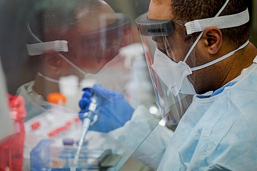
Cytotoxicity Testing for Medical Devices
Methods - using Brine Shrimp
What is Cytotoxicity? - Essentially, cytotoxicity refers to the ability of cytotoxic agents to destroy living cells. Cytotoxic compounds (e.g., immune cells and some venoms) can achieve this by either inducing necrosis or apoptosis.
Cytotoxicity Tests - In many laboratories, cytotoxicity tests are important assays used for the purposes of assessing the cytotoxic potential of various devices and materials.
From these tests, it becomes possible to determine whether given devices/materials are biocompatible for actual use. This is especially important given that some devices or materials have the capacity to impede cell growth or even induce cell death.
Cytotoxicity Testing for Medical Devices
A number of methods are used for testing the cytotoxicity of medical devices.
These methods are divided into three main categories that include:
Direct cytotoxicity test - This includes tests that analyze the effects of devices on cell growth within the same medium. Here, the device being tested is placed in a medium with cells to determine whether it stops the growth of the cells or causes them to die.
Indirect cytotoxicity tests - Here, scientists analyze the cytotoxic effects of any substances leaching out of the device being investigated to determine whether they diffuse through the agar and cause harm the cells.
Extract tests - In this test, scientists add extractable substances from the device to a medium to study their impact on the cells.
Direct Cytotoxicity Test
Also known as the direct contact method, this approach is generally used for plastics and chemical material rather than high-density material. This is due to the fact that unlike plastics, high-density molecular materials have been shown to cause physical cellular damage when they come in direct contact with the cells during the test.
Some of the cells that have often been used for this method are L-929 mouse fibroblasts. Here, however, preparing several plates is recommended for control purposes. Cells are first introduced into the dish/container with complete culture medium for growth.
Following the introduction of the cell into the medium, they are first allowed to adhere to the bottom of the container (e.g., petri dish) and incubated at 37 degrees C with about 5 percent carbon dioxide.
Once the cells grow to about 80 percent confluency, the material to be tested is then placed directly onto the cell in order to determine how it affects the cells.
To test the cytotoxicity of the material, the relatively low-density material is placed directly on the cells and incubated for about 24 hours. If the material under investigation is a liquid (e.g., chemical), then a piece of filter paper (sterilized) is first saturated with the material before the saturated filter paper is directly placed on the cells.
This is then incubated for about 24 hours at about 37 degrees C. After 24 hours of incubation, the cells are examined under the microscope in order to determine whether the material had any cytotoxic effects on the cells.
The grade of reactivity (cytotoxic effect) ranges from 0 to 4 depending on the effects of the material/device on the cells.
* As mentioned, relatively low-density materials are often used in direct contact method. This is because these materials interact well with the cells in the culture. In a case where very low-density materials are used; they tend to float in the medium.
As well, very high-density material can cause physical damage to the cells thus affecting the quality of results
* One of the biggest advantages of this method is that it's highly sensitive and thus allows researchers to identify low cytotoxicity grades of a material.
* For the control sample, the material under investigation is not added to the plate. As a result, the sample without the material/device under investigation can be compared to the samples where it was introduced to the cultured cells.
Indirect Contact Method (Indirect Contact Cytotoxicity Test)
Whereas direct contact method involves bringing the material under investigation in direct contact with cultured cells in the medium, indirect contact cytotoxicity tests are generally used for leachable materials. These are materials that can diffuse through various barriers to reach the cells.
In this case, then, the materials/devices are not brought in direct contact with cultured cells. Rather, a barrier is placed between the cells and the material so that researchers can determine whether any materials leached/diffused through to reach and affect the cells.
There are several types of indirect contact methods which include:
Agar Diffusion Test (Agarose Overlay Test)
Agar diffusion cytotoxicity test is similar to direct contact method in that the material under investigation is introduced into the dish with the cells being grown. The main difference between the two methods is that unlike direct contact method, the agar diffusion test involves using agar which protects the cells from dense materials/devices.
As mentioned, relatively low-density materials are used in direct contact method. For the agar diffusion test, high-density materials (e.g., elastomeric closures) can be used given that the agar serves to cushion the cells from the material/device under investigation.
Although agar is used for this technique, cells to be used have to be cultured in the appropriate medium first. This allows the cells to grow before the material/device is introduced.
Following the initial culture, a layer of agar, which also contains nutrients required for growth, is added to the cultured cells. This creates a thin layer of agar on the cultured cells. The layer of agar has to be allowed to solidify before the test sample (material or device) is placed on top of the agar.
When the thin layer of nutrient-supplemented agar solidifies, the material under investigation can then be placed on top of the agar and incubated. Here, extracts from the material can also be used on a filter paper.
While the agar acts as a cushion/barrier between the cells and the material under investigation, it's sufficiently permeable to allow various substances (gases, toxic compounds, and nutrients, etc.) to leach/diffuse through.
In cases where neutral red dye is also used, studies have shown toxic materials, from the material or devices, along with the dye to be internalized into the cell and transported to the lysosomes.
When they cause the lysosome to rupture, the dye forms a halo in the culture which is an indication that the material contained toxic substances. As well, the degree of destruction caused to the cells can be observed under the microscope.
Some of the main advantages of agar overlay test include:
- It's cheap
- It's simple and can be completed within a short period of time
- Can be used for a wide variety of medical devices/materials
* Although it has several advantages, one of the main disadvantages of this method is that the agar used as a barrier does not properly represent biological barriers in the body.
Molecular Filtration
Molecular filtration is also a type of indirect contact test. Here, however, the aim is to determine the impact of the sample under investigation on the metabolic activity of cells.
As is the case with the other tests, cells used in the test are first cultured. First, the monolayer of cells are grown in a filter of cellulose ester.
As the cells continue growing, the medium is gradually replaced with nutrient supplemented agar so that fresh medium gel is introduced to the cells. Ultimately, the gel, which contains a single layer of cells is separated and reversed so that the cells are exposed to the sample (device under investigation). The filter is then removed before measuring the metabolic activities of the cells.
In particular, molecular filtration seeks to measure the impacts of medical devices on succinate dehydrogenase, which is one of the enzymes found in the mitochondria.
By affecting the metabolic activities of a cell, some medical devices can stop the cells from growing or cause them to die. This method is therefore especially important for investigating this particular impact of cytotoxic devices on the cells.
Some of the main advantages of molecular filtration include:
- Can be used to investigate the primary and secondary cytotoxicity of medical devices and material
- Simple and only takes a short period of time to complete
- Has been shown to be highly sensitive and reliable
Extract Cytotoxicity Test
Extract cytotoxicity test is one of the three types of tests used to test the cytotoxicity of medical devices. In particular, this method is commonly used to test the toxicity of soluble substances from medical devices.
For this method, the materials under investigation are first extracted in serum with minimal essential medium (MEM e.g., serum-supplemented mammalian cell culture media) at about 37 degrees C. The extracts are then placed onto a monolayer of cells so that the cells are allowed to grow in the extraction fluid.
Although cells are allowed to grow in the extraction fluid, it's worth noting that cells to be used for this test are usually first grown in the appropriate culture.
For instance, cells may first be cultured and maintained in Dulbecco's modified Eagle’s medium consisting of fetal bovine serum and 1% penicillin-streptomycin to deter the growth of pathogens. The cells are then incubated at 37 degrees C and about 5 percent carbon dioxide before they are ultimately added to the extraction fluid.
Following the growth of cells in the extraction fluid, they are inspected to determine the effects of the device/material extracts. Here, assessment of the cells may involve the use of a microscope to investigate any morphological changes of cell lysis. This is known as the qualitative approach and cytotoxicity is graded between 0 to 4 depending on severity.
Generally, devices or medical materials with a cytotoxic score of below 2 are said to pass the test. Apart from microscopy, tetrazolium dye can also be used to analyze the impact of the device/material on the metabolic activity of the cells.
If more than 70 percent of the cells remain viable, the material/device is said to pass the test.
Cytotoxicity Testing using Brine Shrimp
Also known as brine shrimp lethality bioassay, cytotoxicity testing using brine shrimp involves the use of brine shrimp (larvae) to test the toxicity of various materials (heavy metals and various compounds etc.).
Procedure
For this method, the material suspected of having cytotoxic effects is first prepared. This involves preparing different concentrations of the sample in order to determine how different concentrations of the material will affect the shrimp.
This may involve the following few steps:
Measure about 10 mg of the sample and dissolve in 1 liter of water - This is the stock solution
Through serial dilution, prepare four (4) different concentrations of the sample; 1mg/mL, 100ug/mL, 10ug/mL, and 1ug/mL
Following the dilution process, equal amounts of the solution are then added to different test tubes, and the larval stage of brine shrimp introduced into the tubes in order to expose them to different concentrations of the material.
To determine the impact of the material on the nauplii (larval stage of brine shrimp), the number of surviving larvae is counted after 24 hours. Here, it becomes possible to not only determine whether the material/substance was toxic, but also the toxicity of different concentrations of the material.
* Freshly hatched brine shrimp can survive for a period of 48 hours without food. During this period, they survive on their yolk sac. For this reason, using freshly hatched brine shrimp (nauplii) is recommended because their death would be attributed to the material rather than starvation. However, yeast can also be used to feed the nauplii 24 hours after hatching.
* While cytotoxicity testing using brine shrimp is an effective method of analyzing the toxicity of various materials, it's important to ensure that the right solvents are used during sample preparation. This is because some solvents are toxic and capable of killing the organism.
In this case, the results would be false-positive because the death of the organisms may be as a result of the solvent rather than the material under investigation.
Some of the main advantages of this method include:
· Is simple and the process only takes a short period of time to yield results
· Has low requirements
· While a large number of organisms are used for statistical validity, the method is inexpensive
See also: Cell Proliferation and Cell Cycle, Fluorescent Dyes in Microscopy
Return from Cytotoxicity Testing to MicroscopeMaster home
References
Chao Wu. (2014). An important player in brine shrimp lethality bioassay: The solvent.
Girish Kumar Srivastava et al. (2018). Comparison between direct contact and extract exposure methods for PFO cytotoxicity evaluation.
Quazi Sahely Sarah, Fatema Chowdhury Anny, and Mir Misbahuddin. (2017). Brine shrimp lethality assay.
Weijia Li, Jing Zhou, and Yuyin Xu. (2015). Study of the in vitro cytotoxicity testing of medical devices.
Links
https://www.eurofins.com/medical-device/services/biocompatibility-testing/cytotoxicity/
https://www.mddionline.com/testing/practical-guide-iso-10993-5-cytotoxicity
https://info.gbiosciences.com/blog/bid/164400/what-is-cell-cytotoxicity-and-how-to-measure-it
Find out how to advertise on MicroscopeMaster!


