Lysosomes
Types, Morphology, Function, Process and Microscopy
What are Lysosomes?
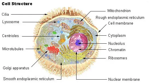
Lysosomes are the main digestive compartment of the cell. As such, they contain a variety of enzymes capable of degrading different types of biological material including nucleic acids, lipids and proteins among others.
They can be found in animal cells and some plant cells (occurring as vacuoles) and are capable of breaking down various types of macromolecules brought in to the cell to be degraded. Most of these are either damaged or have completed their life cycle and are no longer useful.
In addition to these macromolecules, lysosomes also serve to break down cells once they die. While they can be found in almost all cells in animals (except red blood cells) they are particularly abundant in tissues/organs that are involved in high enzymatic reactions.
These include such tissues/organs as the liver, kidney, macrophages and pancreas among a few others. Cells of these tissues/organs contain abundant lysosomes.
* The name lysosome originated from Greek words Lysis (meaning destroy/dissolve) and Soma (meaning body).
* Animal cells may contain numerous lysosomes (several hundred) plant and yeast cells typically have a single, large lysosome (vacuole).
Types of Lysosomes
There are two main types, these include:
Primary lysosomes - are formed from Golgi apparatus appearing as small vesicles. Although primary lysosomes are popular on Golgi apparatus, they also occur as granulocytes and monocytes. These lysosomes are surrounded by a single phospholipid layer and contain acid hydrolases.
The pH value of the acid in these vesicles is important in that its changes activate or deactivate the enzymes. Ultimately, most of the primary granules will fuse with phagosomes, which results in the formation of secondary lysosomes.
Secondary lysosomes - are formed when primary lysosomes fuse with phagosomes/pinosome (they are also referred to a endosomes). The fusion also causes the previously inactive enzymes to be activated and capable of digesting such biomolecules as nucleic acids and lipids among others.
Compared to primary lysosomes, secondary are larger in size and capable of releasing their content (enzymes) outside the cells where they degrade foreign material.
A majority of lysosomal enzymes function inside the acidic environment, which is why they are referred to a acid hydrolases.
They contain about 45 enzymes that are grouped in to six main categories:
- Nucleases - Nucleases are important enzymes that hydrolyze nucleic acids. Nucleases are divided in to deoxyribonuclease (acts on DNA) and ribonuclease which hydrolyses RNA. Hydrolysis action on nucleic acids results in the production of sugars, nitrogen bases as well as phosphates.
- Proteases - Proteases includes enzymes like collagenase and peptidases that acts on proteins converting them to amino acids
- Glycosidases - Glycosidases like beta galactosidase act on the glycosidic bonds of polysaccharides converting polysaccharides to monosaccharides. For instance, the enzyme galactosidase acts on such bonds converting lactose to glucose and galactose.
- Phosphatases - Good examples of Phosphatases are acid phosphodiesterases. These are important enzymes that act on organic compounds releaing phosphate in the process. However, the compound has to have a phosphate group.
- Lipases - Lipases include esterases and phospholipiases that act on lipids to produce acids and alcohol
- Sulphatases - Sulphatases are enzymes that act on organic compounds to release sulphates
* Lysosomes cannot digest themselves - Most of the proteins present in its membrane contain high amounts of carbohydrate-sugar groups. Because of the present of these groups, digestive enzymes are unable to digest the proteins present on the membrane.
Morphology/Structure
Lysosomes are membrane-delimited organelles. This means that they are surrounded by a membrane that prevents its components from being released. This is particularly important given that uncontrolled release of the acidic fluid and enzymes can cause damage to the components of the cell.
They also have a high concentration of protons, which results in pH value of less than 5.
The surrounding membrane is composed of integral proteins as well as a vacuolar-type H+ ATPase, highly glycosylated proteins and a number of transporters. Depending on the type of lysosome and their function, they also greatly vary in size (between 1 micrometer and several microns) and general shape.
They are less defined compared to other types of organelles. When viewed, they appear as cytoplasmic dense bodies that may be ovoid, spheric or tubular on occasion.
Function
The manner in which lysosomes function highly depends on the way the enzymes affect other materials outside and inside the cell. There are a number of processes through which lysosomes digest material.
These include:
Endocytosis
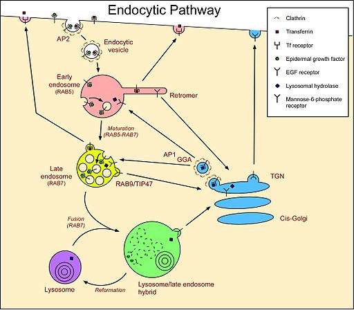
Endocytosis is one of the most popular phenomenon exhibited by cells. In endocytosis, invagination of the plasma membrane of the cell results in the creation of an endocytic vesicle that engulfs different types of extracellular molecules.
In phagocytosis (a type of endocytosis) large molecules/microorganisms like bacteria are engulfed in a vacuole (phagosome). However, in pinosomes (another type of endocytosis) a small amount of the surrounding fluid and solute molecules are pinched off as pinosomes (pinocytic vesicle).
Once these vesicles fuse with primary lysosomes, secondary lysosomes are formed. Enzymes are then activated and act on the molecules. This process is commonly referred to as the "Endosomal Pathway"
See also: Endocytosis Vs Exocytosis
* Digested material is typically passed into cellular component while undigested ones are excreted.
Process
Material uptake- formation of endosome - formation of the phagolysosome - lysis- diffusion of digested material and exocytosis.
Autophagic Lysosome
Apart from endocytosis, lysosomes are also involved in another process referred to as autophagocytosis. This process helps in the degradation of various cell components that are either worn out or malfunctioning.
In addition to simply breaking down cellular components, this process helps in recycling of these material. Here, autophagic lysosomes (secondary) release enzymes that digest various cell components as soon as the cell dies. This is also referred to as autolysis.
Autophagy also takes place during starvation. During starvation periods, lysosomes will start hydrolyzing organic foods that are stored in cells so as to produce energy.
Vacuoles in Plants
In plants, vacuoles serve many functions, which make them multifunctional organelles. However, they also have basic properties similar to lysosomes found in the cells of animal. As such, they have an acidic nature and contain various hydrolic enzymes capable of breaking down different types of molecules.
There are different types of vacuoles that serve different functions. These include:
- Sap vacuoles - these vacuoles store salt minerals and nutrients
- Contractile vacuoles - involved in osmoregulation and excretion
- Food vacuoles - involved in the digestion of nutrients - this vacuoles contain digestive enzymes that digest these nutrients
- Air vacuoles - serve to store metabolic gases in prokaryotes
Differences and Similarities between Primary and Secondary Lysosomes
Similarities
- Both have acid hydrolases
- Both have a single phospholipid membrane surrounding them
Differences
- Whereas primary lysosomes are buds that form from Golgi apparatus, the secondary are formed from fusion of primary lysosomes and phagosomes or pinosomes
- Primary lysosomes largely serve to store material (as storage vacuoles) while secondary largely serve as digestive vacuoles where lysis occurs through the activity of hydrolytic enzymes
- While the primary do not release any of their content, secondary lysosomes can release their content (enzymes) in to the cell cytoplasm for exocytosis. Exocytosis is where enzymes are released in to the cytoplasm or outside the cell where they degrade different types of material.
- For most part, primary lysosomes have inactive hydrolases while secondary have active lysosomes capable of acting on molecules and other material
Recycling
Lysosomes are also important in that they act as recycling centers. Whenever different types of molecules or cell components are broken down (for instance proteins broken down to amino acids) the amino acids are then used as building blocks of new proteins. This ensures that some of the byproducts are re-used in the body.
On the other hand, they help recycle material that are not easy to excrete. A good example of this is iron. When iron is released from the breakdown of various cells or cell components (such as red blood cells), iron is recycled and used for the construction of new organelles. This allows for a minimized excretion of the by-products and retention of others to be used in the body.
Microscopy
Lysosomes are too small to view using a light microscope. For this reason, an electron microscope is used to observe them. However, it is possible to view a lysosome (vacuole) in a plant cell.
The following is a procedure to view plant vacuole:
Requirements
- An onion
- Glycerin
- Safraning solution
- A pair of forceps
- A dropper
- Microscope glass slides
- A light compound microscope
- Distilled water
- Microscope cover slip
- Two watch glasses
Procedure
- Pour a few drops of distilled water in to a watch glass
- Using a pair of forceps, peel off a membrane from the skin of an onion and place it in the watch glass with a drop of water
- Add a few drops of safranin in to the other empty watch glass
- Pick the onion membrane using the forceps and place it in the watch glass with safranin and allow it to stand for about 30 seconds
- Pick the membrane again and return it in to the watch glass with distilled water
- Add a drop of glycerin in to the center of a microscope glass slide
- Place the onion membrane on the glass slide (in the glycerin) and cover with a cover slip
- Place the slide under the microscope and observe
Observation
Apart from viewing many irregular cells and a cell nucleus, students will clearly see a large vacuole at the center of the cell.
An overview of the different organelles
Do animal cells have vesicles?
Differences between cytosol and cytoplasm
Take a look at the Nucleus, Mitochondria, Ribosomes, Golgi Apparatus , Vacuoles
Return from Lysosomes to Cell Biology
Return to MicroscopeMaster Home
References
Vinay Kumar (2012) Complete Biology for Medical College Entrance Examination.
Links
http://www.sciencedirect.com/science/article/pii/S1357272511002366
Find out how to advertise on MicroscopeMaster!


