Sensory Cells
Characteristics, Location, Function and Microscopy
Definition: What are Sensory Cells?
Commonly referred to as sensory neurons, sensory cells are specialized cells capable of sensing and distinguishing information (any changes in the external environment) through sensory receptors present on their surface. As such, sensory cells play an important role in receiving and relaying information from the external environment that allows the central nervous system to register the stimuli and initiate the appropriate response.
As front-liners in closest proximity to the stimuli, sensory cells allow the organism to respond appropriately to such stimulus as light, sound, taste, temperature, and pressure among others. The body has different types that are classified based on the morphology, location and specific function (the stimulus that the cell reacts to).
Given that sensory cells play a role in conducting information relating to touch, smell, taste, and auditory inputs among others, they respond to impulse generated from the following sensory organs:
- Eyes
- Nose
- Tongue
- Skin
- Ears
Characteristics
While different types of sensory cells exist, which have different functions with regards to the stimuli they react to; they share a number of characteristics between them.
Despite being located in different regions of the body, sensory cells are activated by input (stimuli) from the external (as well as some internal) environment. While these inputs (stimuli) may vary (physical or chemical), sensory cells, as front-liners in the nervous system, receive and send this information for further processing and response.
Although sensory cells have different types of receptors that are the first to come into contact with the stimulus, these receptors are generally divided into two main categories that include exteroceptors and interoceptors. Whereas interoceptors respond to changes inside the body (muscle tension, blood pressure, etc), exteroceptors sense stimuli from the external environment (cold, heat, pressure, etc).
Depending on the type and function, sensory cells are also classified according to the structure of the sensory receptors. For instance, while some of the cells have receptors with free nerve endings, others have encapsulated nerve endings with enclosed terminal ends. Regardless, information received from these receptors (physical or chemical stimuli) has to be converted to electrical impulses that are transmitted to the central nervous system.
Location and Function of Sensory Cells
As previously mentioned, sensory neurons cells that sense different types of stimuli from the external (as well as internal) environment relay that to the central nervous system. As such, they make up the afferent system, which obtain information from the sensory organs (skin, etc).
These sensory cells extend to different parts of the body and to the sensory epithelium (which are concentrated with such sensory cells as the olfactory sensory neurons). This allows them to collect information from the external environment and relay that to the central nervous system.
In the sensory epithelium, sensory cells are located in different regions and vary in morphology. This allows them to effectively sense the appropriate stimuli and relay the information.
Sensory receptors (also referred to as sensory receptor cells in some books) are structures of the sensory cells that are embedded in the sensory epithelium where they collect information from the external and internal environment.
Different types of receptors include:
- Chemoreceptors
- Pain receptors
- Thermoreceptors
- Mechanoreceptors
- Photoreceptors
While sensory receptors are located in different sensory epithelia that allow them to collect the appropriate information (with changes in the environment), they are also widely classified based on their complexity.
While some of the receptors have a special capsule that encloses the nerve ending, commonly known as encapsulated receptors, others lack this structure and therefore have free nerve endings.
Location
Exteroceptors
Exteroceptors include a group of sensory receptors that detect any changes from the external environment. As such, they are well positioned to respond to various stimuli that come from outside the body. This includes such stimuli as vision, temperature changes, touch, smell and pain among others.
There are four types of exteroceptors which include:
Mechanoreceptors
Mechanoreceptors include a group of receptors located under the skin. These receptors respond to such physical changes as touch, vibration, stretch and pressure among others.
Some of the receptors identified as mechanoreceptors include:
- Pacinian corpuscles
- Merkel complexes
- Meissner corpuscles
- Ruffini corpuscles
Pacinian corpuscles
Compared to some of the other mechanoreceptors, Pacinian corpuscles are located deeper in the dermis.
With regards to structure, Pacinian corpuscles have an onion-like capsule where a fluid-filled space separates the inner core and outer membrane lamellae. They also have a myelinated nerve ending with an outer layer made up of flattened cells, a lymph-like fluid, and collagen fibers.
They fall under the category of cells that have encapsulated endings. As compared to Meissner's corpuscles, the Pacinian corpuscles tend to adapt faster and therefore have a lower response threshold.
Pacinian corpuscles are involved in the detection of pressure and vibrations that are transient in nature.
Some of the other characteristics of Pacinian corpuscles include:
· They have one or several rapidly afferent axons that originate from the onion-like capsule
· Their nerve endings are activated by transient disturbances that range between 250 and 350 Hz
· Stimulation of their afferent fibers produces a sensation of vibration or tickle
· In the hand, they make up about 15 percent of the cutaneous receptors
Merkel complexes
Unlike Pacinian corpuscles, Merkel complexes/Merkel's disks are located in the epidermis and thus aligned with the papillae fund in the dermal ridges.
Merkel's disks are also slow adapting, as compared to Pacinian corpuscles. They are more in numbers making up about 25 percent of the total mechanoreceptors of the hand. For the most part, they are found in higher concentration in the lips, external genitalia, and fingertips. In the epidermis, these receptors respond to light pressure.
* Merkel complexes are classified under free nerve endings receptors.
Meissner’s corpuscles
Meissner’s corpuscles/tactile corpuscles are located in the upper epidermis. However, they can also be found projecting into the epidermis of the fingertips, palms, eyelids, and soles.
With regards to structure, Meissner's corpuscles are elongated and composed of lamella or Schwann cells. Like Pacinian corpuscles, Meissner's corpuscles are fast adapting.
Unlike the other receptors, however, Meissner's corpuscles make up the majority of mechanoreceptors in the skin contributing to about 40 percent of the sensory innervation in the hand. Being encapsulated endings, Meissner's corpuscles are well able to respond to fine touch and pressure as well as vibrations of low frequencies.
Ruffini corpuscles
Also known as bulbous corpuscles, Ruffini corpuscles are mechanoreceptors that are located in the hairy and glabrous skin. As slow adapting and encapsulated receptors, Ruffini endings are located in the stretch lines of the skin where they detect any stretching (of the digits or movement) or deformations.
Pain receptors (nociceptors)
Also known as nociceptors, pain receptors are located in the skin (epithelial layer) and other tissues. These receptors respond to a variety of noxious stimuli resulting from mechanical, chemical and thermal factors.
There are several types of pain receptors (peripheral nociceptors) which include:
· Thermal nociceptors - Respond to changes in temperature (above 52 and low 5 degrees Celsius): These receptors are located in the skin and the viscera.
· Polymodal nociceptors - Respond to such pain as those caused by heat and algesic chemicals: These receptors are located in the skin and joints.
· Silent nociceptors - respond to such proinflammatory mediators like histamine and serotonin: These receptors are located in the joints and viscera
· Mechanoheat nociceptors - Respond to changes in temperature and pressure: These receptors are located in the skin, viscera, and joints.
· Visceral nociceptors - Located in such regions as the serosa surface and intestinal muscle and respond to such pain as those caused by stretching and contraction of the viscera.
In different parts of the body, these receptors are responsive to such pain from:
- Cutting and burning (on the skin)
- Spasms and stretching of the mesentery (in the gut)
- Rupture, tears, and swells (of the skeletal muscle)
- Joint inflammation
Compared to most of the other receptors which respond to low sensitivities, nociceptors have been shown to only respond to stimuli causing tissue damage. Therefore, in the event that these receptors are stimulated, a certain part of the body has experienced some level of tissue damage.
* Pain receptors are examples of receptors that lack any special structures (unencapsulated).
Proprioceptors
Proprioceptors are a type of mechanoreceptors that sense information regarding angle of the joint, muscle tension as well as muscle length. As such, they provide information about limb position in space.
Two of the most important Proprioceptors include:
Muscle spindle - Enclosed in a capsule, muscle spindle are tiny sensory organs found in muscle. Within the body of the muscle, this receptor provides information regarding changes in the length of the muscle.
Golgi tendon organ - Unlike muscle spindles, Golgi tendon organ is found in the tendons where they provide information regarding changes in muscle tension as a result of a tendon pull.
Thermoreceptors
Thermoreceptors are divided into cold and warm thermoreceptors.
Whereas cold receptors detect any changes that fall below body temperature, warm thermoreceptors detect changes in temperature that goes above the normal body temperature.
Some of the receptors capable of detecting changes in temperature include:
· Krause end bulbs - Detect temperature changes below body temperature.
· Ruffini endings - Ruffini endings are a type of thermoreceptor that can detect temperatures above body temperature.
Another type of thermoreceptor with free nerve endings can detect temperature changes (cold and warm).
These receptors can be found in such body regions as the liver, skin, and the skeletal muscle. Information from these regions is transmitted through the A-delta and C-fibers and transported to the central nervous system.
Although they are both important, in that they help detect changes in temperature, studies have shown the cold thermoreceptors to account for the majority of these receptors.
* Generally, thermoreceptors are free, non-specialized nerve endings. However, some may have a very thin capsule.
Chemoreceptors
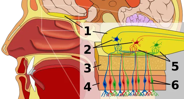
Chemoreceptors include such receptors as taste buds (located on the tongue) and olfactory cells (located in the sensory epithelium of the nasal cavity).
In their respective locations, these receptors detect taste and odors thereby making it possible to for such primary tastes as sweet, bitter, sour and salty (through the taste buds) as well as a range of odorants that enter the nose.
The two are specialized and contain proteins that bind to chemicals from the external environment causing them to become depolarized, which in turn results in an action potential.
Apart from the two chemoreceptors, which detect chemicals from the external environment, for the most part, the body also contains such internal chemoreceptors as the peripheral chemoreceptor (that detect changes in pH in the aortic and carotid bodies) and the central chemoreceptor in the brain that responds to changes of pH in the cerebrospinal fluid.
Sensory Cells Microscopy
Requirements:
- Sample: Sensory epithelium - E.g sensory epithelium obtained from the labyrinth of animals
- Phase contrast microscope
- Alcohol
- Xylol
- Canada balsam
Procedure:
Samples used for this procedure should not have been stored for more than a week given that doing so tends to affect the integrity of the tissue.
- Using the alcohol, dehydrate the specimen - this is used to remove water from the sample
- Use xylol to dissect the specimen
- Use Canada balsam to mount the specimen
- Place the sample under phase contrast microscope for observation
Observation:
When viewed under the microscope, it's possible to see some elongated cells with terminal parts of myelin sheaths which are dark in color.
Return to Cell Biology main page
Return to learning about Nerve Cells
Return from Sensory Cells to MicroscopeMaster home
References
Bruce Alberts, Alexander Johnson, Julian Lewis, Martin Raff, Keith Roberts, and Peter
Henrik Henriksön Lindeman. (2013). Studies on the Morphology of the Sensory Regions of the Vestibular Apparatus
Min‐Chuan Huang. Receptors and Signal transduction.
Natasa Popovic. (2010). Cell Surface Receptors. ResearchGate.
Seol Hee Im and Paul H. Taghert. (2011). PDF Receptor Expression Reveals Direct Interactions between Circadian Oscillators in Drosophila. NCBI.
Walter. (2002). Molecular Biology of the Cell. 4th edition.
Sven Dijkgraaf. (2019). Mechanoreception.
Links
https://www.dartmouth.edu/~rswenson/NeuroSci/chapter_3.html
https://opentextbc.ca/biology/chapter/17-3-taste-and-smell/
Find out how to advertise on MicroscopeMaster!

![Three Basic Types of Neuronal Arrangements-Artwork by Holly Fischer [CC BY 3.0 (https://creativecommons.org/licenses/by/3.0)] Three Basic Types of Neuronal Arrangements-Artwork by Holly Fischer [CC BY 3.0 (https://creativecommons.org/licenses/by/3.0)]](https://www.microscopemaster.com/images/256px-Three_Basic_Types_of_Neuronal_Arrangements.png)
![Dorsal Root Ganglion by OpenStax [CC BY 4.0 (https://creativecommons.org/licenses/by/4.0)] Dorsal Root Ganglion by OpenStax [CC BY 4.0 (https://creativecommons.org/licenses/by/4.0)]](https://www.microscopemaster.com/images/1318b_Dorsal_Root_Ganglion.jpg)


