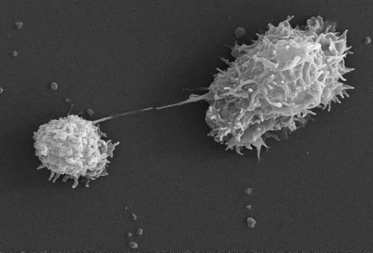
Amoeba Under The Microscope
Fixing, Staining Techniques and Structure
Amoeba (plural amoebas/amoebae) is a genus that belongs to Kingdom protozoa. Generally, the term is used to describe single celled organisms that move in a primitive crawling manner (by using temporary "false feet" known as pseudopods).
Amoebas are eukaryotes, which means that their genetic material are well organized and enclosed within a membrane (nuclear membrane)
* The word amoeba comes from the Greek word "amoibe" which means to change
Amoeba Microscopy
Amoebas are simply single celled organisms. As such, they can only be viewed using a microscope. There are methods that can be used to observe these organisms.
The first and simplest methods involves viewing amoebas under the microscope without staining. This is a simple method that allows students to view them live as they move around. The second method involves fixing and staining to get a better view of the structure and organelles of the organism.
1. Simple (Direct) Method
Amoebas can be found freely living and thriving in shallow pond waters with organic material.
To view amoebas under the microscope, students will need the following:
- A sample of water collected from a pond with organic material
- Pondweed from a pond
- Petri dish
- A compound light microscope
- Water
- A dropper
For this technique, the student may either observe a sample of pond water directly to identify the organism or conduct a simple culture to grow and increase the number of amoebas.
Amoeba culture is a simple exercise that involves the following few steps:
- Place a few pondweeds in a petri-dish and add some water to cover the weed
- Leave the culture is a dark room for a few days and wait until a brown scum forms on the surface
Procedure for Microscopy
- Using a dropper, place a few drops of the sample on a microscope glass slide (a sample of pond water or a small sample from the culture)
- Gently cover the sample with a cover slip and mount on the microscope stage for viewing
- Start with low power and increase gradually to observe the specimen
Observation
When viewed, amoebas will appear like a colorless (transparent) jelly moving across the field very slowly as they change shape. As it changes its shape, it will be seen protruding long, finger like projections (drawn and withdrawn).
2. Fixing and Staining Technique
Requirements
- Amoeba culture
- A bench-top MSE centrifuge
- Neff's saline, sea water
- Moist chamber
- Cover slip
- Sodium hydroxide (NaOH)
- Distilled water
- Nissenbaum's fixative
- Lugol's Iodine
- Formalin seawater
- Carnoy's fixative
- Heidenhain’s iron Haematoxylin
For this technique, the specimen (amoebae) is first cultured using such culture and saline agar slopes. Following the culture, a sample of the specimen is further concentrated using a centrifuge (at 3 Krpm for about 10 minutes).
This is continued by the following few steps:
- Wash the pellet using 75 percent seawater, Neff's saline or any other appropriate solution
- Allow the specimen to settle on a cover slip in a moist chamber until the amoebae adopt a normal locomotionary morphology (the cover slip can be treated with sodium hydroxide depending on the type of amoeba)
Fixing
- While settling on the cover slip, pipette freshly prepared Nissenbaum’s Fixative on the sample and allow to stand for 5 minutes
- Wash the sample with acidified HgCl2 for about 7 minutes
- Wash the sample with 50%, 35%, 15% ethanol for about 5 minutes
- Wash the sample again with distilled water for about 5 minutes
Some of the other fixatives that can be used include:
- Carnoy’s Fixative
- Lugol’s Iodine
- Seawater formalin
Staining
Staining the amoebae is aimed at enhancing visibility of the mitotic figures.
Some of the stains that can be used to stain the amoeba cells include:
- Heidenhain’s iron Haematoxylin
- Kernechtrot (Nuclear Red)
- Modified Field’s Stain
- Klein’s Silver relief stain
For Heidenhain’s iron Haematoxylin, staining involves the following steps:
- Inoculation of the sample in 2 percent ammonium ferric sulphate for about 1 and a half hour
- Rinsing the sample in distilled water
- Inoculation of the sample in 0.5% haematoxylin and 2% ammonium ferric sulphate for about one and a half hours
- Wash the sample with tap water
After staining, mount the slide on the microscope stage to observe.
Observation
When viewed under the microscope, students will notice tiny dark spots in the cytoplasm of the organism while the cytoplasm is lightly stained.
* Direct observation of the organism (without staining) has a great advantage in that the amoebae are still alive and motile when being viewed under the microscope.
This allows the students to see the finger like projections (pseudopods) elongate and shorten as the organism moves about or engulfs given substrates. However, this technique does not allow students to view the cell’s organelles.
Fixing and staining on the other hand kills the amoeba, which means that students will not get to see the organism moving in the field of view but staining increases contrast, allowing students to get a better view of the organelles in the cell.
Structure of Amoeba
As mentioned, amoebae are eukaryotes, which simply means that they have a cell membrane surrounding their cytoplasm and DNA that is properly packed in the central nucleus.
When viewed under the microscope, these aspects of the organism are clearly visible particularly when the sample is stained.
Some of the other organelles that are visible under the microscope
include:
- A food vacuole
- Cytoplasm
- Contractile vacuole
See differences between cytosol and cytoplasm here.
* With regards to their structure, amoebae closely resemble cells of higher animals like those of human beings.
Pseudopodia
Essentially, Pseudopodia are temporary projections of the cytoplasm that make it possible for amoebae to move. Pseudopods are some of the most distinguishable features of amoebae and their formation is based on the flow of the protoplasm.
The organism contracts in a manner that pushes the cytoplasm to fill and expand a pseudopod while pulling at adhesions at the back of the cell.
According to studies, the process
involves the following steps:
- With pressure from ectoplasm, which is an exterior gel, endoplasm (interior fluid) is forced to flow forwards in the cell.
- On reaching the tip of the membrane, the pressure causes the endoplasm to form a pseudopod.
- The endoplasm is then forced back towards the ectoplasm and turns to gel (this causes the new pseudopod to disappear) The new gel is again converted to endoplasm and again under pressure moves to the membrane to form a new pseudopod.
Apart from using pseudopod to move around, amoebae also use them to engulf food particles. Here, the pseudopod surround the particle while an opening on the membrane allows the particle to move into the cell and into a food vacuole where it is digested by enzymes.
Learn about Acanthamoeba and Naegleria Fowleri
Return to Pond Water under the Microscope
Return to Microscope Experiments Page
Return from Amoeba under the Microscope to Microscopy Research home
Sources
Kwang Jeon (1973) Biology Of Amoeba.
Links
http://www.scienceclarified.com/Al-As/Amoeba.html
http://www.bms.ed.ac.uk/research/others/smaciver/Staining_amoebae.htm
Find out how to advertise on MicroscopeMaster!
![Amoeba Proteus by Joseph Leidy (Fresh-water Rhizopods of North America, 1879) [Public domain], via Wikimedia Commons Amoeba Proteus by Joseph Leidy (Fresh-water Rhizopods of North America, 1879) [Public domain], via Wikimedia Commons](https://www.microscopemaster.com/images/AmoebaproteusfromLeidy.jpg)
![Amoeba diagram by el:User:Kupirijo (Amoeba_(PSF).png) [GFDL (http://www.gnu.org/copyleft/fdl.html) or CC-BY-SA-3.0 (http://creativecommons.org/licenses/by-sa/3.0/)], via Wikimedia Commons Amoeba diagram by el:User:Kupirijo (Amoeba_(PSF).png) [GFDL (http://www.gnu.org/copyleft/fdl.html) or CC-BY-SA-3.0 (http://creativecommons.org/licenses/by-sa/3.0/)], via Wikimedia Commons](https://www.microscopemaster.com/images/Amoebadiagram.png)


