Naegleria Fowleri Amoeba
Characteristics, Symptoms, Treatment and Infection
Overview/Introduction
A member of the phylum Percolozoa, Naegleria fowleri is an environmental ameboflagellate commonly found in warm water and terrestrial environments (aquarium, ponds, swimming pools, etc).
While it belongs to a group of free-living organisms, Naegleria fowleri is pathogenic and a causal agent of primary amebic meningoencephalitis (an infection of the central nervous system) in human beings.
Although these infections are rare, they are fatal with a 98 percent death rate. Currently, infections caused by the organism have been identified in less than 20 countries. However, its distribution is largely limited to environmental conditions in which the cyst can survive.
Some of the symptoms associated with primary amebic meningoencephalitis include:
- Fever
- Vomiting
- Headache
- Impaired mental status
* Naegleria fowleri is named after Dr. Malcolm Fowler, an Australian pathologist who (along with Dr. Rod Carter) recognized the organism as a causal agent of primary amebic encephalitis in 1965s.
Classification of Naegleria fowleri
Kingdom: Protista - As members of the kingdom Protista, Naegleria fowleri are simple eukaryotic organisms commonly found in various terrestrial and water bodies where they exist as free-living organisms or as parasites/pathogens capable of causing disease. As members of this kingdom, they are not plants, animals or fungus.
Subkingdom: Protozoa - Members of the subkingdom Protozoa, e.g. Naegleria fowleri, are eukaryotic single-celled organisms that can be found all over the environment where they exist as free-living organisms or as parasites. They vary highly in shape with some of the species being capable of changing their shape.
Phylum: Percolozoa - Members of the phylum Percolozoa colorless amoebae that are characterized by flagellate phases in their life cycle. The majority of the organisms (amoeboflagellates) are free-living and can be found in various aquatic and terrestrial habitats where they feed on bacteria and other organic material.
Class: Heterolobosea - Acarpomyxea is a class that consists of several orders including: Schizopyrenida. Members of the class can be found in soil, marine, and freshwater environments where they live as free-living organisms. Some of the species, however, can be parasitic and cause diseases/infections in human beings.
Order: Schizopyrenida - Members of the Order Schizopyrenida are naturally occurring in soil and aquatic habitats. The majority of species in this group are free-living in these environments and thus do not directly depend on a host.
Family: Vahlkampfiidae - This family includes such genera as Naegleria, Monopylocystis, and Paravahlkampfia etc which inhabit various aquatic and terrestrial habitats as free-living organisms (the majority of organisms are free-living). Members of this group are often referred to as limax amoebae most of which are characterized by flagella.
Genus: Naegleria - The genus Naegleria consists of over 45 species. Members of this group are free-living protists commonly found in warm water bodies and some terrestrial habitats across the globe.
Of these species, only N. fowleri and N. lovaniensis are known to be pathogenic with N. fowleri being the only species known to cause disease in human beings. In addition to being naked amoebae, members of this group are also characterized by their transformation from amoebae to flagellates.
Species: Naegleria fowleri - Characteristics of this species are discussed below.
Ecology
As free-living organisms, the species Naegleria fowleri can be found in diverse terrestrial and aquatic habitats across the world (except the Antarctica). Apart from the soil and aquatic habitats, Naegleria fowleri has also been isolated from the air under certain conditions.
In parts of Nigeria, for instance, the amoeba has been isolated from the air during the Harmattan (a season characterized by dry and dusty winds).
In aquatic environments/water bodies, such as freshwater lakes, population of the organism has been shown to vary with changes in temperature. While the number is relatively high during the hot months of summer, the population peaks late in the summer. Here, however, it's worth noting that different temperature ranges favor different forms of the amoeba.
While trophozoites thrive in a temperature range between 35 and 46 degrees C, flagellates require temperatures of between 27 and 37 degrees C. Apart from warm freshwater lakes, some of the other aquatic habitats in which Naegleria fowleri have been isolated include ponds, rivers, swimming pools, waste (sewage), thermally polluted water, and contaminated water, and hot springs.
* In their habitats, Naegleria fowleri can survive pH range of between 2.1 and 8.15.
* While N. fowleri can be found in swimming pools, it cannot survive in chlorinated pools. Low temperatures (cool) and clean water also affect the ability of the organism to reproduce and multiply.
Apart from various water bodies, Naegleria fowleri can also be found in soil at temperatures over 30 degrees C. However, they are commonly found in moist or wet soil that allows flagellate forms to swim in water films.
Soil is particularly an important habitat for the organism given that it provides some conditions that promote growth and multiplication. These include oxygen, favorable temperature ranges, and bacteria (food source).
From the soil, the amoeba can then be transported to various water bodies by streams or heavy rains, etc. It's from these sources (water bodies) that the organism can infect human beings.
* Naegleria fowleri cysts can survive adverse environmental conditions such as low temperature ranges.
* Healthy human beings are also carriers of N. fowleri - The organism has been detected in swabs obtained from the mouth, nose, and pharynx of health patients as well as human sera and saliva samples.
Morphology and Cell Structure of Naegleria fowleri
The life cycle of Naegleria fowleri is characterized by three distinct morphological stages. Therefore, in order to describe the general morphology and cell structure of the organism, it's important to look at its life cycle.
Life Cycle
Trophozoites
The life cycle of Naegleria fowleri can occur in various aquatic bodies and terrestrial environments as well as human hosts. This is because they occur as free-living organisms as well as human pathogens.
The life cycle of Naegleria fowleri starts with the trophozoite stage commonly found in aquatic environments. Being the infective stage of the organism found in aquatic environments, the trophozoites can infect human beings who come in contact with the water in infected swimming pools, etc.
While infections are very rare, studies have shown them to affect children and young adults when contaminated water enters the body through the nose.
Trophozoites also represent the reproductive stage of the organism and divide through binary fission under favorable conditions (e.g. temperature range of between 35 and 46 degrees C). This results in the production of two similar cells that can grow and repeat the process.
* During cell division (through binary fission), the nucleolus elongates and divided into two. However, the nuclear membrane remains intact. Karyokinesis, which involves nuclear division, is then followed by division of the cytoplasm (cytokinesis) before the cell ultimately divides into two daughter cells.
Morphology/Structure of Trophozoites
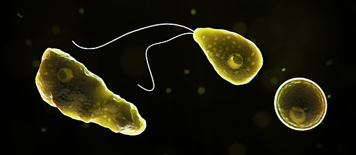
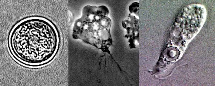
Trophozoites of Naegleria fowleri are small in size, ranging from 10 to 35um in size. They are characterized by a limax-like appearance and have a sticky posterior end that consists of trailing filaments.
Like other members of the genus Naegleria, the trophozoites of Naegleria fowleri exhibit cytoplasmic extensions known as amoebastomes (food cups). These structures may vary in size and number depending on the strain. In aquatic and terrestrial habitats, trophozoites contain a higher number of food cups which are used to ingest such prey as yeast and bacteria as well as for attachment.
As eukaryotic cells, trophozoites of Naegleria fowleri are also characterized by many organelles commonly found in other eukaryotes.
For instance, they have a rounded membrane-bound nucleus that consists of a thick nucleolus, a cell membrane that is about 10nm in width, smooth and rough endoplasmic reticulum, as well as a high number of ribosomes within the cytoplasm among others.
Like many other cells, the trophozoites also have a cytoskeleton structure that supports the cell structure. This consists of thick (located in the cytoplasm) and thin (located near the plasma membrane) microfilaments made up of actin and myosin.
Unlike the flagellate stage, trophozoites use amoeboid locomotion to move from one point to another. Here, movement is made possible by adhesion to various substrates in their surroundings. The speed of movement, however, is largely dependent on temperature.
* The direction of movement of N. fowleri trophozoites is by chemotaxis.
Flagellates
Under favorable environmental conditions, Naegleria fowleri trophozoites reproduce through binary fission to produce two daughter cells. However, in the event of nutritional deficiency and ionic changes, this form of the organism transforms into flagellate forms.
While changes in the environment cause trophozoites to transform into flagellates, this stage still requires water. Unlike trophozoites, flagellates thrive in a temperature range of between 27 and 37 degrees C and cannot reproduce (they also don't eat) - Transformation from the trophozoite stage to flagellate stage only takes a few hours.
Naegleria fowleri flagellates are characterized by a pear-shaped appearance and measure between 10 and 16mm. During this transformation (from trophozoite stage to the flagellate stage), studies have shown the number of vacuoles to decrease as the basal bodies and flagella form.
The flagellum that is ultimately formed has the typical 9 + 2 arrangement that consists of filaments that are surrounded by a sheath. Under favorable conditions, the flagellates can transform back to the trophozoite stage and feed on various prey in their surroundings.
Like the trophozoite stage, Naegleria fowleri flagellates also have a nucleus. However, here, it's located at the anterior region of the organism which is narrower compared to the anterior region. The nucleus, which also consists of a nucleolus, is covered by a distinct nuclear membrane as is the case with the trophozoites.
Some of the other organelles found in the flagellate forms include:
- Mitochondria
- Rough endoplasmic reticulum
- Vacuoles
- Rhizoplast
Cyst Stage
While flagellates are formed in the event of nutritional deficiency or due to ionic changes, the trophozoites transform into the cyst stage in adverse conditions (cold, depleted nutrients, dry conditions, etc).
Like the flagellate stage, cysts are also incapable of reproduction and are also non-feeding. They are spherical in shape and can survive various adverse conditions in which the trophozoites and flagellates cannot survive.
Some of the other characteristics of the Naegleria fowleri cysts include:
- Have a thick wall that has pores
- A mucoid plug seals the pores of the cyst wall
- The encysted cell contains a nucleus and several cytoplasmic vacuoles
- Contain several organelles commonly found on other eukaryotic cells - rough endoplasmic reticulum, and a less pronounced nucleolus
* N. fowleri cysts can also enter the human host through the nasal passages. Once they enter the body, they transform to the trophozoite stages that can then cause an infection.
Metabolism and Pathogenicity
Naegleria fowleri are aerobic heterotrophic organisms commonly found in aquatic and various terrestrial environments (trophozoite forms). As such, they are commonly found in oxygen-rich environments and have many mitochondria.
In these environments, Naegleria fowleri feed on bacteria and other single-celled organisms like yeast. Here, the trophozoites use pseudopods to engulf and ingest food material which are then digested in the vacuoles.
Apart from pseudopodia, N. fowleri also form food-cups on which are phagocytic structures that play an important role in the pathogenicity of the organism.
* Pseudopods extend from the cell surface to engulf the prey.
* A food-cup is formed through the invagination of the body surface.
* According to studies, trophozoites transform into active swimmers (flagellates) when they search for prey. When they arrive in a habitat with sufficient or high amount of prey, they transform back to the trophozoites which are capable of feeding - This is achieved through chemotaxis
Infection
As mentioned, Naegleria fowleri are aerobic heterotrophic organisms commonly found in oxygen-rich habitats. In the body of a human host, the brain is an oxygen-rich environment with conducive conditions that allow the organism to survive as a pathogen.
In humans (particularly children and young adults), acute infections occur when Naegleria fowleri is inhaled through the nasal passages. While the infections are rare, they occur when contaminated water (swimming pool water or other water with trophozoites) or cysts in dust are inhaled through the nasal passages.
* The nasal passage is the only route that allows for successful colonization of the host given that the pathogen can effectively migrate to the brain. If the pathogen is ingested, it's easily destroyed by stomach acids.
Once the organism is inhaled through the nasal passage, the trophozoites attach to the nasal mucosa. Adhesion to the cells of the mucosa is the first and most important step in the pathogenesis of the pathogenic amoeba (made possible by adhesions expressed on the surface of the organism). From here, the amoeba make their way to the olfactory bulbs through the olfactory nerves.
While Naegleria fowleri have been shown to move towards given direction through chemotaxis, the olfactory sensory axon bundles act as tunnels that allow the organism to successfully reach the brain having bypassed the central nervous system barrier.
* If an individual inhales Naegleria fowleri cysts, they first have to transform into trophozoites which are able to attach to the nasal mucosa and migrate to the brain having crossed the porous cribriform plates.
* According to studies, children and young adults are at a higher risk because their cribriform plates are more porous. Located directly posterior to the nares/nostril, the cribriform plates is a porous extension that separates the nasal cavity from the brain.
* Apart from attachment to the nasal mucosa, chemotactic response (with Acetylcholine acting as the chemo-attractant) as well as a high rate of locomotion are some of the other factors that promote pathogenicity of N. fowleri.
In the brain parenchyma, N. fowleri uses such proteins as fibronectin-binding protein to bind to the brain cells. Using the food-cup structures, they start feeding on the brain tissue through a process known as trogocytosis.
Here, the pathogens use their food-cup structures to extract and ingest the cell membrane and intracellular components of the brain cells thus causing extensive damage - This is the reason Naegleria fowleri are also referred to as brain-eating amoeba.
Apart from trogocytosis, Naegleria fowleri also cause damage by releasing cytolytic molecules. Here, however, it's worth noting that different strains of the species affect the host differently. For instance, according to studies, the weakly pathogenic strains tend to cause damage by ingesting brain tissues using the food-cup structures.
On the other hand, strains that are highly pathogenic first lyse the nerve cells before ingesting debris that is produced.
Some of the molecules that have been associated with cell lysis include:
- Proteases
- Acid hydrolases
- Phospholipases
- Phospholipolytic enzymes
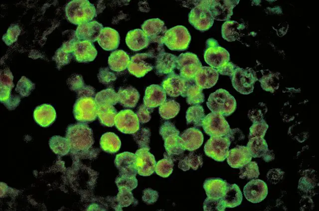
By ingesting brain tissues and releasing various cytolytic molecules, N. fowleri end up causing amebic meningoencephalitis (PAM) which is a serious infection of the central nervous system. It's rapidly occurring (acute fulminating) and may manifest 3 to 10 days following contact with infected water (or after the cyst entered the body through the nasal passage).
Some of the symptoms associated with the infection include:
- Photophobia
- Headache - Bifrontal or bitemporal
- Spiking fever
- Nausea and vomiting
- Stiff neck
- Coma - Coma may ultimately result in death a few days after presenting the symptoms
* N. fowleri has been shown to be resistant to lysis action of the host cytolytic molecules (e.g. tumor necrosis factor etc). This is one of the adaptations that allows it to thrive in the central nervous system.
* While N. fowleri is distributed in different parts of the world, particularly in tropical areas, the majority of infections have been reported in the United States.
Control and Treatment
Naegleria fowleri are sensitive to chlorine and cannot survive in clean water. Therefore, water that has been sufficiently chlorinated (e.g. pool water, etc) as well as properly maintaining water are some of the best strategies to control the organism and highly minimize possible infections.
In the event of an infection, however, Amphotericin B is commonly used for treatment purposes. Although the pathogen is highly sensitive to the drug in vitro, not all patients recover with this treatment.
Some of the other drugs used for treatment purposes include:
- Rifampicin
- Sulphadiazine
- Antiedema medication
Return to Amoeba under a Microscope
Return from Naegleria Fowleri to MicroscopeMaster home
References
Francine Marciano-Cabral. (1988). Biology of Naegleria spp.
Francine Marciano-Cabral and Guy A. Cabral. (2007). The immune response to Naegleria fowleri amoebae and pathogenesis of infection.
Muhammad Jahangeer et al. (2019). Naegleria fowleri : Sources of infection, pathophysiology, diagnosis, and management; a review.
Ruqaiyyah Siddiqui, Ibne Karim M. Ali, Jennifer R. Cope, and Naveed Ahmed Khan. (2016). Biology and Pathogenesis of Naegleria fowleri.
Links
https://web.stanford.edu/group/parasites/ParaSites2010/Katherine_Fero/FeroNaegleriafowleri.htm
Find out how to advertise on MicroscopeMaster!


