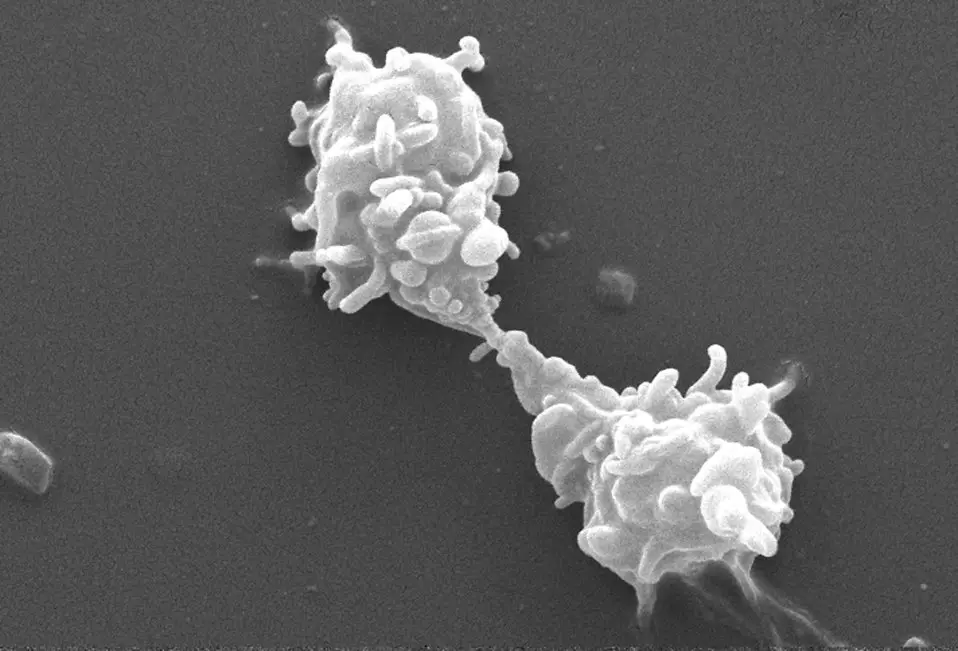
Acanthamoeba
Species, Morphology, Life Cycle, Culture, Disease
Acanthamoeba is a genus of amoebae consisting of a number of species that are pathogenic in nature. They are ubiquitous in nature and are therefore widely distributed across the environment (fresh water, soil, etc).
Examples of include:
- A. polyphaga
- A. astronyxis
- A. rhysodes
- A. lenticulat
- A. divionensis
Protists (Protozoa)
Acanthamoeba belong to the subkingdom protozoa of the kingdom protista. As such, they belong to a group of unicellular organisms known as protists, which are eukaryotic organisms that do not fit in the other eukaryotic kingdoms.
Some of the other eukaryotic kingdoms include:
- Plant kingdom
- Animal Kingdom
- Fungi
* While members of this genus share various characteristics with other eukaryotes, they are not plants, animals or fungi.
Eukaryotes
As eukaryotic organisms, cells of genus Acanthamoeba are organized into complex structures enclosed in a cell membrane. The nucleus of these cells is enclosed in a double layered membrane known as the nuclear envelope similar to that of other higher eukaryotic organisms.
Some of the other membrane bound organelles found in these cells include:
Habitat
(distribution of Acanthamoeba in the environment)
Species of the genus Acanthamoeba have been isolated in a wide variety of environments; both natural and man-made. For instance, while this specie can be found in such natural environments as water ponds, mud and streams (natural environments) they have also been identified in various factory discharges, air-conditioners, cooling towers as well as ventilation ducts among many others.
Apart from such common environments as oceans, beaches and soil, these species have also been isolated in a number of more extreme environments thus making the group an important focus for researchers.
One of the best examples of this was the isolation of these organisms in the Antarctica; an environment with extremely low temperatures and dry winds. This discovery proved that the organisms are present in virtually all environments across the world.
Members of this genus have also been found in:
- Dead and decaying animals
- Droppings of such birds as pigeons
- The digestive system of some reptiles
Morphology/Structure
Studies have identified more than 20 species of genus Acanthamoeba that are divided into three main groups based on their size and cyst morphology.
These include:
Group 1 Species
Species classified as group 1 are characterized by a large cyst (over 18um) and rounded ectocyst. Trophozoites of group 1 Acanthamoebas are also larger in size and have been shown to range from 25 to 35 um in size. While the cyst size has played an important role of identifying group 1, they have also been associated with the following characteristics:
They form ectocysts (outer wall) and endocysts (inner walls) that have a clear separation
Group 1 species are largely environmental organisms and cause little human and animal infections compared to the other groups.
Group 1 consists of 5 species including:
- A. astronyxis
- A. echinulata
- A. tubiashi
- A. comandoni
Group 2 Species
Group 2 consists of about 10 species. Compared to Group 1 species, species of Acanthamoeba in Group 2 have a smaller cyst (and smaller trophozoites) while the endocyst varies in shape.
Unlike Group 1 species, this Group has also been shown to cause a majority of human infections.
Some of the other characteristics of this Group include:
· The endocyst and ectocyst may be closer compared to those of Group 1 or clearly separated
· The ectocyst may appear triangulate, round or polygonal in shape
· Cysts of Group 2 specie are less than 18um in size
Examples of Group 2 species include:
- A. castellannii
- A. quina
- A. triangularis
- A. rhysodes
- A. hatchetti
- A. divionensis
- A. griffini
Group 3 Species
Compared to Group 1 species, species that belong to Group 3 are also smaller in size.
5 species have been classified in this group and they include:
- A. culberlsoni
- A. pustules
- A. palestinensis
- A. royreba
- A. lenticulata
Genotypes
While its size and morphology have played an important role in the identification and classification of these organisms, recent studies have been able to group these species based on the sequence of their genomic DNA.
To date, about 17 genotypes of Acanthamoeba have been identified. This has proved to be a more effective way of identifying various species compared to using morphological features for identification purposes: The use of morphological features has been shown to be problematic due to inconsistencies and variants of the cyst morphology.
Some of its genotypes that have been clearly identified include, T1, T2a (and T2b), T3, T4*. T5, T6, T7, T8, T9, T10, T11, T12, T13, T14, T15.
Genomic sequencing has shown there to be a 5 percent variance in the genomic sequence of these genotypes, making this a more effective method of identifying the different genotypes. These small differences between the genotypes (including the 4.9 percent variance between T2a and T2b) have made this method more effective and thus the method of choice for many researchers.
While many species have been shown to cause various opportunistic infections, Acanthamoeba keratitis infection (AK) and other non-keratitis infections have largely been associated with T4. These are serious infections that not only affect sight, but also various other organs in the body.
* T4 is associated with about 90 percent of all keratitis infections. It is also associated with such non-keratitis infections as Acanthamoeba granulomatous encephalitis and other skin infections.
|
Genotype and Strains |
Organ Infected |
T1
Acanthamoeba sp. strain CDC V006
Acanthamoeba sp. strain OSU 03-022/CDC V329
Brain
T4
Acanthamoeba sp. strain CDC V168
Acanthamoeba sp. strain CDC V328
Acanthamoeba sp. strain OSU 03-009/CDC 12741:1
Skin
Brain
Lungs
Acanthamoeba sp. strain OSU 03-021/CDC V313
Nasal
Other strains like Acanthamoeba sp. strain OSU 03-029/CDC V501 and Acanthamoeba sp. strain OSU 03-030/CDC V503 (belonging to T4) have been isolated in the cerebrospinal fluid, which shows that they can affect the central nervous system.
Life Cycle
This genus goes through two main life stages that include a trophozoite stage (active stage) and the dormant stage where cysts are formed.
Trophozoite Stage
This trophozoite stage is the vegetative and motile phase that occurs during favorable conditions (neutral pH, availability of sources of nutrition, optimal temperature- about 30 degrees etc).
Depending on the genotype, generation time ranges from 8 to 24 hours as the cells passe through the growth cycle. During this phase, the organisms actively feed on a variety of organic substrate particles as well as other unicellular organisms such as yeast and bacteria.
* The trophozoite stage is the infective phase
* While they feed on various single celled organisms, it has also been shown to act as a reservoir for bacteria. According to studies, such bacteria as Mycobacterium avium, not only live in Acanthamoeba (as they are transported to the next host) but also multiply in these organisms.
Cyst Stage
During the cyst stage, Acanthamoeba form a cyst, which allows the organisms to survive tough environmental conditions. For instance, the cyst is capable of surviving lack of food sources, desiccation, extreme pH and temperatures as well as ultraviolet irradiation among others.
Growth Cycle
In favorable conditions, Acanthamoeba cells divide asexually through a process known as binary fission. This process produces two identical daughter cells that continues the life cycle.
The growth cycle is divided into the following phases:
· Lag phase- This is the first phase in which the cells take time to adapt to their environment. It is also during this stage that the Acanthamoeba cells start maturing - Some of the molecules synthesized as the cells mature include enzymes and RNA.
· Exponential phase- With favorable conditions available (nutrients, temperature etc) the number of cells condition to increase in the exponential phase. The cells continue to double until nutrients are depleted (and waste/toxins material increase). Depletion of nutrients and the buildup of toxins lead to the next phase in the cycle.
· Stationary phase- The stationary phase is the result of high number of cells from the exponential phase, depletion of nutrients and increase of toxins. These conditions result in reduced rates of growth
· Cell death and encystation- As a result of depleted nutrients and high levels of toxins, the cells start to die or form cysts to ensure their survival. Cyst formation allows the cells to survive in various extreme conditions by remaining dormant until environment conditions improve.
One of the other methods of describing its life cycle involves dividing the phases into two main stages.
These include:
Interphase - The interphase period is divided into the following phases:
- G1 - In the G1 phase, various molecules including enzymes are synthesized as the cells prepare for the next phase of growth. Some of the cells start to divide in this phase.
- S - DNA synthesis and replication takes place in the S phase.
- G2 - During this phase, DNA synthesized in the S phase contributes to high protein synthesis to produce microtubules. About 90 percent of cell division also takes place during this phase.
* Microtubules produced during the G2 phase are important for mitosis (cell division) that occurs in the M phase
M phase - Nuclear division occurs in the M phase. Chromosomes are separated to form two daughter nuclei in preparation of cell division. While nuclear division (as well as cytoplasmic division) occurs during this phase, the M phase only accounts for about 10 percent of cell division in the growth cycle.
Acanthamoeba Cell Biology
As mentioned, the life cycle of Acanthamoeba goes through two main stages (trophozoite and cysts). Being eukaryotic cells, the trophozoites contain various organelles that are present in higher animals and plants including a prominent nucleus, ribosome, mitochondria and a Golgi apparatus among others.
In addition to these organelles, these cells also exhibit acanthopodia (pseudopodia), which are spike-like structures used for movement and trapping prey. The pseudopodia of these organisms are capable of adhering to various surfaces, which makes it possible for the cells to move and even capture prey.
* Acanthamoeba use their acanthapodia to trap and capture various particles including single celled organisms like bacteria among other organisms to feed on. These particles are consumed through a process known as phagocytosis (or pinocytosis in some cases). Here, the surface of the cell invaginates and ultimately results in the particles being digested by various enzymes in the food vacuole.
Infections
Acanthamoeba has been associated with a number of infections in both human beings and animals. However, some of the most common infections include:
Acanthamoeba Keratitis
Acanthamoeba keratitis is a serious infection of the eye that can result in blindness if not attended to. While it is rare, the infection has been shown to mostly affect those who wear contact lenses for a long period of time. However, it can also affect those who do not use such lenses in cases of poor personal hygiene.
Once the organism attaches to the cornea of the eye, it starts to breakdown the epithelial layer while depleting the keratocyte following stromal invasion.
If left untreated, the infection can ultimately cause stromal necrosis that can in turn cause visual impairment or complete blindness.
Acanthamoeba Granulomatous Encephalitis (AGE)
Also referred to as Granulomatous Amebic Encephalitis, AGE is a serious infection of the brain and the spinal cord that typically affects those with a poor immune system.
In such cases, Acanthamoeba has been shown to enter through the respiratory system and ultimately reach the brain and spinal cord fluid through blood circulation. Because the immune system of the individual is compromised, the organisms are successfully transported to these organs where they can cause edema and hemorrhagic necrosis.
Treatment
Early intervention has been shown to be successful in the treatment of these infections. Whereas Acanthamoeba keratitis is treated using such drugs as chlorhexidine digluconate in combination with hexamidine or propamidine, the treatment or management of Acanthamoeba granulomatous encephalitis involves using such drugs as sulfadiazine, ketoconazole, itraconazole and amphotericin B among others.
Culture
Requirements
- Non-nutrient agar
- Page's Saline
- Incubator
- Agar plates
- Pipette
- Centrifuge
- Test-tubes
- Sample
- E. Coli
Procedure
- Add the non-nutrient agar in to two agar plates (clean sterile plates) and incubate at 35 degrees Celsius for 30 minutes
- Using a tube of Page's saline, prepare a dense suspension of E. coli
- Using a clean pipette, add a few drops of the suspension into the agar plates (2 to 3 drops)
- Spread the suspension on the agar plate to ensure even distribution
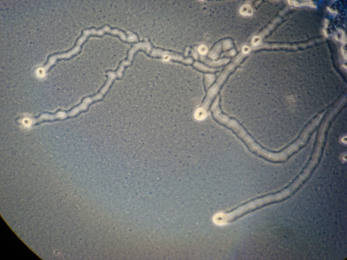
* E. coli agar allows for Acanthamoeba to be detected through feeding tracks in the media
Sample preparation (of the Acanthamoeba)
Samples may be obtained from different sources including, water bodies, soil, cerebral spinal fluid and contact lenses among others. Depending on the source, the sample is prepared differently. This section will focus on soil sample preparation.
Procedure
- Add about 2 grams of the sample into a tube of Page's Saline
- Using a pipette, add a few drops of the suspension onto the agar plates (E. coli agar plates)
- Incubate the plates for about 24 hours at about 35ºC
- Seal the plates using paraffin strips and continue incubating (invert the plates)
- Observe the plates under the inverted microscope
Observation
Feeding tracks in the plates indicates the presence of Acanthamoeba
Image – This image shows feeding tracks in the agar plate, which indicates the presence of Acanthamoeba (A. castellanii)
Significance of Acanthamoeba
While this specie causes various infections in human beings and some animals, they have been shown to play an important role in controlling the population of certain micro-organisms in the environment as well as a role in the regulation of various nutrients and elements in soil and water bodies.
Return to page on Amoebas under the Microscope.
Return from Acanthamoeba to MicroscopeMaster Home
References
Naveed Ahmed Khan. Acanthamoeba: Biology and Pathogenesis. Originally published: 2009.
Naveed Ahmed Khan. Acanthamoeba : biologyand increasing importance in human health. School of Biological and Chemical Sciences, Birkbeck College, University of London, London, UK. Received 11 October 2005; revised 9 March.
Ruqaiyyah Siddiqui and Naveed Ahmed Khan. Biology and pathogenesis of Acanthamoeba. Parasites & Vectors 2012, 5:6
Links
https://en.vircell.com/diseases/21-acanthamoeba-castellanii/
https://catalog.hardydiagnostics.com/cp_prod/Content/hugo/FreeLivingAmebaeMedia.html
Find out how to advertise on MicroscopeMaster!


