Chlamydia Bacteria
Classification, Characteristics and Microscopy
What is Chlamydia?
Chlamydia bacteria is a genus that is obligatory intracellular. Currently, three species of Chlamydia responsible for human infections have been identified with each having one or several serovars. Although they are responsible for a number of infectious diseases in human beings, they also cause numerous infections in other animals (birds, pigs, cattle, horses, etc).
As causative agents of various diseases in both human beings and animals, the Chlamydia bacteria species are prevalent worldwide (in both industrialized and developing countries) with varying infections depending on the species. In the United States and Australia, for instance, Chlamydia trachomatis is one of the most common bacterial agent of STDs (sexually transmitted diseases).
Examples of Chlamydia species include:
- Chlamydia suis
- Chlamydia trachomatis
- Chlamydia psittaci
- Chlamydia avium
- Chlamydia abortus
- Chlamydia pecorum
* Species responsible for human infections include Chlamydia trachomatis, Chlamydia pneumoniae, and Chlamydia psittacci.
Classification of Chlamydia
Domain: Bacteria - As bacteria, Chlamydia bacteria are prokaryotic cells. As such, they have a simple cell structure lacking membrane-bound organelles. Bacteria can be found in various environments across the world, existing as either free-living organisms or as parasites.
Phylum: Chlamydiae - Chlamydiae is a phylum and class that consists of obligate intracellular bacteria. Members of this group have been shown to be highly diverse and infect both human beings and animals. However, some species do not cause harm to their host and live as symbionts. Generally, members of the phylum Chlamydiae are oval in shape and are Gram-negative.
Order: Chlamydiales - Bacteria classified under the order Chlamydiales are obligate intracellular organisms that are known to cause human and animal infections. According to recent studies, some of these pathogens have been shown to be transmitted by ticks (which act as vectors). As such, they are able to infect vertebrates, invertebrates as well as free-living amoebae in some cases.
Family: Chlamydiaceae - Chlamydiaceae is a family that consists of Gram-negative bacteria of the phylum Chlamydiae. It only consists of a single genus that in turn contains a number of species that infect both human beings and animals
Genus: Chlamydia
Characteristics
Distribution
According to research studies, the family Chlamydiaceae, which consists of the genus Chlamydia originated from the Order Chlamydiales seven million years ago. However, infections associated with Chlamydia trachomatis have been traced back to the 27th Century BC, causing sexually transmitted diseases and chronic ocular disease.
Today, Chlamydia bacteria are distributed all across the world with C. trachomatis being the most sexually transmitted infection among young adults.
While Chlamydia species are widely distributed across the world, the rate of infections vary from one region to another. For instance, while Chlamydial STD infections are widely distributed across the world, blinding trachoma has been shown to be particularly predominant in parts of Africa, Asia, and Western Pacific with high rates of poverty.
In 2004, the prevalence and distribution of Chlamydia bacteria were as follows:
· About 89 million (92 million in 2002) new cases of sexually transmitted Chlamydia infections globally
· Prevalence of 4.19 percent among young people in the United States (18 to 26 years of age)
· 6 million cases of irreversible trachoma blinding with 146 million requiring treatment
· Half of the adult population above the age of 20 (worldwide) exhibit serological evidence of past C. pneumoniae infections
· C. trachoma accounts for about 15 percent of blindness across the world
Cell Biology
Members of the genus Chlamydia are Gram-negative bacteria. While the majority of these bacteria have a thin peptidoglycan, the peptidoglycan of these species is not easily detectable.
In C. pneumoniae and C. trachomatis, however, genomic studies have shown that they encode for proteins that may be involved in the synthesis of peptidoglycan. In addition, they encode for a major outer membrane protein, a protein located on the surface of C. trachomatis and C. psittaci. This is an important protein used for serological identification of different forms of C. psittaci and C. trachomatis.
* The absence of peptidoglycan in Chlamydia bacteria has been suggested to be an evolutional adaptation that allows them to effectively invade and thrive in the cells of their respective hosts.
* Infective and reproductive forms of Chlamydia include elementary bodies (EB) and reticulate bodies.
Some of the other morphological characteristics of Chlamydia bacteria include:
Chlamydia trachomatis
- not motile - lack structures required for movement
- shape varies from coccoid to rod-shaped
- reticulate bodies range from 800 to 900nm in diameter while the infectious bodies (elementary bodies) range from 300 to 400nm in diameter
Chlamydia pneumoniae
- They exhibit a coccoid shape which may be elongated
- Elementary bodies are pear-shaped and range from 200 to 400nm in diameter
Reproduction and Life Cycle of Chlamydia
As mentioned, there are three species of Chlamydia responsible for human infections. These include C. trachomatis, C. pneumoniae, and C. psittacci. However, there are different ways in which each of the species infect human beings.
Whereas C. trachomatis is sexually transmitted from one human being to another, C. psittaci, which infects birds, can be transmitted through contact with such birds as parrots causing parrot fever. In human beings, these infections are mostly asymptomatic which can contribute to their spread.
The incubation period has also been shown to vary between strains within these species. While this may take a few days in some strains of C. trachomatis, it can take months to several years in the strains of C. pneumoniae.
Chlamydia Bacteria infections also cause different diseases/illnesses in human beings: C. psittaci may cause pneumonia or mild illnesses, C. trachomatis are responsible for sexually transmitted diseases, while C. pneumoniae is responsible for infections in the upper and lower respiratory tract.
Diseases caused by Chlamydia can also be divided based on the mode of transmission. There are two main categories that include transmission through direct contact and transmission through the respiratory route. In the case of inclusion conjunctivitis, trachoma, or lymphogranuloma, the transmission of C. trachomatis may occur through contaminated fingers (urogenital tract to eyes), using a contaminated towel and through an infected birth canal in neonates.
In genital infections, however, this bacteria is transmitted through C. trachomatis. C. psittaci and C. pneumoniae are transmitted through the respiratory tract. In the case of C. psittaci, this transmission is from infected birds to human beings while the transmission occurs from one infected human being to another in the case of C. pneumoniae.
In their life cycle as pathogens, Chlamydia bacteria alternate between two morphologically distinct forms; elementary body and the reticulate body. During infection, cell invasion is by the elementary bodies, which are metabolically inert. These forms are extra-cellular and have to invade the cell in order to give rise to the reticulate forms.
Alternation of Elementary and Reticulate Bodies in the Life Cycle of Chlamydia
Elementary body (EBs) are metabolically inert and small in size. They are characterized by a rigid outer membrane that consists of proteins with a high content of disulfide cross-linked cysteine.
This membrane is beneficial for the parasite given that it allows them to survive the harsh conditions of the extracellular environment. In order to penetrate the cells of the host, the elementary body first attaches to their cell membrane using their ligands. These allow the bacteria to attach to the receptors of the host before they can inject their effectors into the cytosol of the cell (of the host).
While the entire mechanism of this attachment is not fully understood, a number of host proteins are also suspected to contribute to this attachment. These include the mannose 6-phosphate receptor, platelet-derived growth factor receptor, as well as the estrogen receptor complex among others.
Following the injection of pre-packaged effectors into the cytosol of the host, an inclusion (vacuole) is formed housing the elementary bodies as they transform into reticulate bodies.
In some cases, many elementary bodies bind to the cell membrane of the same cell (of the host) resulting in the formation of many inclusions. These inclusions may fuse to form a single, large inclusion in which the parasites reside as they transform to form the reticulate bodies.
* Within the inclusions, it takes about two hours for elementary bodies to transform into reticulate bodies.
* Whereas the elementary bodies have a membrane that protects them in the extra-cellular environment, reticulate bodies encapsulate themselves in a cytoplasmic vacuole which allows them to evade cellular lysosomes of the host. This behavior is suggested to protect them from antibiotics.
The reticulate bodies formed in the inclusions are metabolically active and tend to be strictly intracellular. They are also larger compared to the elementary bodies with the largest ones having a diameter of about 1um.
Reticulate bodies are characterized by a diffuse and fibrillar nucleic acid-rich granular cytoplasm. Unlike elementary bodies, reticulate bodies are not infectious and consist of an inner and outer membrane. Moreover, they are covered by projections and rosettes on their surface which may project through the membrane of the inclusion.
Within the inclusions, reticulate bodies undergo binary fission within several hours which allows them to rapidly increase in numbers. In the process, reticulate bodies give rise to elementary bodies that are released into the extracellular environment 48 to 72 hours after the initial infections.
This cycle allows Chlamydia bacteria to infect and destroy new cells depending on how the bacteria exit the cell. Once the parasite is transmitted to another person (through the means of transmission mentioned above) this cycle also continues.
Mechanism by which Elementary Bodies exit the Host Cell
Following the transformation of reticulate bodies into elementary bodies, they are released from the cell (of the host) in one of two ways; lysis of the host cell or extrusion of the inclusion (vacuole).
Lysis of the host cell is destructive and results in the death of the cell. The process starts with the rapture of the membrane (inclusion membrane) by cysteine proceases. This is then followed by rapture of the plasma membrane of the host cell to release the elementary bodies. This process causes the permeabilization of both the intrusion membrane and nuclear membrane and ultimately the lysis of the plasma membrane - This is calcium-dependent.
Extrusion of the inclusion follows the interaction between the cell of the host and the bacteria. This contact results in the invagination of the vacuole (vacuole that contains the Chlamydia) followed by furrowing of the plasma membrane of the host cell. This process results in the pinching of the cell which expels the inclusion from the host cell. Generally, this process leaves the host cell intact.
Microscopy of Chlamydia Bacteria
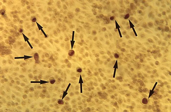
In clinical settings, Chlamydia bacteria infections can be detected through microscopy. This method is particularly used to investigate the presence of inclusions in infected cells.
Requirements
- Sample
- Microscope - Fluorescent microscope
- 95 percent ethanol
- Clean glass slide
- Giemsa stain
Procedure
Sample Collection
Chlamydia bacteria samples are often collected using cotton, Dacron, and calcium alginate swabs from an infected patient. Here, swabs with wooden shafts are not recommended given that the wooden shaft may be toxic which in turn affects the specimen.
Depending on the infection, the sample may be obtained from the ocular or genital tract, anal canal, nasal etc. If the sample is not to be prepared immediately for microscopy, then it has to be stored under conducive conditions. For instance, nasopharyngeal swabs are placed in cell culture media.
Staining Procedure
· Create a smear of the sample on a clean glass slide - This can be achieved by simply smearing the swab on the slide
· Allow the smear to air dry
· Fix the specimen using absolute methanol for about 5 minutes and allow it to air dry
· Cover the specimen with Giemsa (the stain should be freshly prepared) for about 1 hour
· Rinse the slide rapidly in 95 percent ethanol - This step serves to remove excess dye
· Observe the slide under a fluorescent microscope
Observation
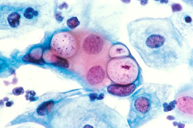
When observed under the microscope, the inclusions will appear pinkish-blue in color (because they are basophilic). The presence of inclusions in the sample is indicative of Chlamydia infection.
Return from Chlamydia bacteria to MicroscopeMaster home
References
Arlieke Gitsels, Niek Sanders and Daisy Vanrompay. (2019). Chlamydial Infection From Outside to Inside.
Cherilyn Elwell, Kathleen Mirrashidi, and Joanne Engel. (2016). Chlamydia cell biology and pathogenesis.
Georg Häcker. (2018). Biology of Chlamydia.
Wandy L. Bea1ty, Richard P. Morrison, and Gerald I. Byrne. (1994). Persistent Chlamydiae: from Cell Culture to a Paradigm for Chlamydial Pathogenesis.
Zuck, Meghan, Stephens, Richard, Hybiske, Kevin. (2017). Contribution of the Exit Mechanism Extrusion in Chlamydia trachomatis Pathogenesis.
Links
https://www.sciencedirect.com/topics/immunology-and-microbiology/chlamydia
Find out how to advertise on MicroscopeMaster!


