Glycosomes
Structure, Function and Location
Definition: What are Glycosomes?
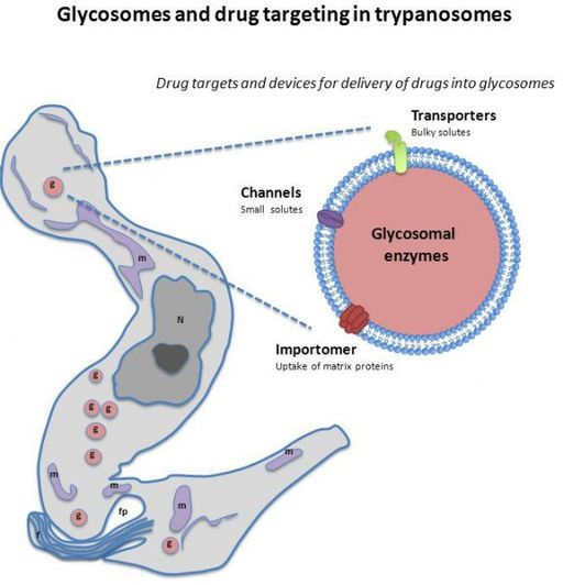
Glycosomes are membrane-bound microbodies of the family peroxisomes commonly found in kinetoplastids and diplonemids. While biosynthesis and metabolic processes within glycosomes are similar to those of microbodies found in other eukaryotic organisms, they also contain additional enzymes required for such processes as glycerol metabolism and gluconeogenesis among others.
A few examples of enzymes found in the glycosome include:
- Hexokinase
- Phosphofructokinase
- Catalase
* Peroxisomes (also known as microbodies) are membrane-bound cytoplasmic organelles involved in oxidative functions.
Structure and Characteristics of Glycosomes
The total number of glycosomes per cell is largely dependent on the species and development stage of the organism.Whereas the bloodstream form of T. brucei may contain between 60 and 65 glycosomes, this number may increase to over 120 during growth and cell division.
According to other studies, the number of glycosomes per cell may range from 80 to 340 during the different development stages of the parasite. With regard to size and general appearance, glycosomes have been shown to be mostly homogeneous.
* Glycosomes of T. brucei are about 0.27um in diameter.
Like peroxisomes found in other eukaryotic cells, glycosomes are membrane-bound organelles surrounded by a single membrane consisting of phosphatidylcholine and phosphatidylethanolamine. In addition, they have an electron-dense proteinaceous matrix but lack any DNA.
Glycolytic enzymes present in the organelles make up a significant amount of the proteins with other enzymes (enzymes involved in such processes as peroxide metabolism and the oxidation of fatty-acids etc) making up a small portion of these molecules.
Given that glycosomes lack DNA, protein synthesis occurs in the cytosol (within the cytosol of free ribosome) and transported to the organelles. Here, the proteins may be transported to the glycosome as either fully folded proteins or oligomeric complexes.
In such organisms as T. brucei, the total content of protein in the organelle is about 150mg/ml and has been associated with crystalloid inclusions within the microbodies. Glycosome composition has also been shown to change at different developmental stages (life cycle stages).
For instance, in the bloodstream forms of various kinetoplastids, ATP is normally generated through glycolysis with about 90 percent of glycosomal protein content being involved in this process. However, generation of ATP in procyclic forms of the parasite occurs in the presence of glucose (of using amino acids in the absence of glucose).
This difference is largely due to the fact that among procyclic forms, the mitochondrion is well developed and fully functional.
* The compartmentation of various glycosome enzymes is not only important for metabolism purposes but also survival of the organism. Based on a number of studies, it has been shown that the expression of enzymes like phosphoglycerate kinase in the cytosol kills the parasite.
Glycosome Functions
As compared to other eukaryotes where glycolysis occurs in the cytosol, enzymes involved in glycolysis are compartmentalized in the glycosome in kinetoplastids. For this reason, the organelle is essential for the survival of the organism given that it is the site of several metabolic processes.
Some of these processes include:
Glycolysis
Like a number of other parasites, the life cycle of kinetoplastid parasites is dependent on two hosts (vertebrates and invertebrates). In both hosts, metabolism of the parasite has to adapt in order for the parasite to not only continue developing through differentiation but also survive.
In the mid-gut of the insect (invertebrate host), the parasite uses mitochondrial metabolism due to the presence of amino acids, threonine, and glutamic acid, etc. Here, this type of metabolism is essential for these food sources.
In the bloodstream, however, parasitic forms are exposed to a glucose-rich environment that requires the glycolytic pathway for metabolism. As a result, mitochondrial metabolism is suppressed in favor of glycolytic metabolism that is restricted in the glycosomes.
Here, the process starts with the conversion of a glucose molecule into glucose-6-phosphate that is then converted to fructose-1, 6-diphosphate. Through a series of reactions, the diphosphate is then converted to glyceraldehyde-3-phosphate, PEP, and pyruvate.
It may be converted to dihydroxyacetone phosphate which is, in turn, converted to glycerol-3-phosphate in the presence of NADH. Ultimately, the process produces two moles of pyruvate and ATP from a mole of glucose.
Gluconeogenesis
As already mentioned, the mid-gut of the insect hold has limited glucose. For this reason, parasitic forms in this environment have to use amino acids available in order to generate glucose.
Although the manner in which fluxes of glycolysis and gluconeogenesis is regulated is not properly understood, studies have shown that the process (gluconeogenesis) is carried out through phosphoenolpyruvate carboxykinase and fructose-1,6-bisphosphatase enzymes located in the glycosomes of the insect stage of the parasite.
Here, it's the tight regulation of both glycolytic and gluconeogenic fluxes that ultimately generate glucose required for energy production.
Pentosephosphate shunt and Oxidation of Fatty Acids
In glycosomes, various enzymes are responsible for the metabolism of sugars like ribose and erythrose which are C5 and C4 sugars. On the other hand, they contribute to lipid metabolism.
C5 and C4 sugars are produced through pentose-phosphate shunt from glucose 6-phosphate with glucose-6-phosphate dehydrogenase and 6-phosphogluconate dehydrogenase playing a central role in the detoxification of reactive oxygen species produced in the organelle.
While the oxidation of fatty acids is yet to be fully understood in glycosomes, studies have shown that beta-oxidation enzymes associated with the organelle may play an important role in the process.
Nucleotide Metabolism
Given that some kinetoplastids are unable to synthesize purines, they rely on some glycosomal enzymes involved in the interconversion of host nucleosides into purine nucleotides in the purine salvage pathway.
Although some of the enzymes, like dihydroorotate dehydrogenase, are located in the cytosol, pyrimidine salvage enzymes uracil phosphoribosyltransferase and uridine-specific nucleoside hydrolase as well as Orotate phosphoribosyltransferase and ornithine decarboxylase which are also involved in the pathway are located in the glycosomes.
During this process (purine salvage), purine made in this organelle is then exported to be used in the nucleic acid of the organism.
Some of the other functions of glycosomes include:
· Synthesis of sterol - through the isoprenoid biosynthetic pathway
· Ether-lipid synthesis - some of the enzymes involved in ether-lipid synthesis include alkyl DHAP synthase and DHAP acyltransferase. Found in the glycosomes, these enzymes carry peroxisome targeting signal.
Take a look at the Pentose Phosphate Pathway as well.
See the parasite: Trypanosoma
Return to Organelles main page
Return to Parasites under the Microscope
Return to Kinetoplastid parasites
Return from Glycosomes to MicroscopeMaster home
References
Fred Opperdoes. (2010). The Glycosome of Trypanosomatids.
Juliette Moyersoen, Jungwoo Choe, Erkang Fan, Wim G.J. Hol and Paul A.M. Michels. (2004). Biogenesis of peroxisomes and glycosomes: trypanosomatid glycosome assembly is a promising new drug target.
Sarah Bauer and Meredith T. Morris. (2017). Glycosome biogenesis in trypanosomes and the de novo dilemma.
Links
https://www.frontiersin.org/articles/10.3389/fcimb.2020.00025/full
Find out how to advertise on MicroscopeMaster!


