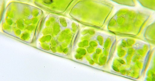
Chloroplasts
** Definition, Structure, Function and Microscopy
What are Chloroplasts?
Essentially, chloroplasts are plastids found in cells of higher plants (plants with advanced traits with lignified tissue for transport of water and minerals) and algae as sites of photosynthesis. This makes them the most important cell organelles given that plants are the primary producers and the base of all food chains.
Depending on the type of plant or algae, the number of chloroplasts in a cell may range from 1 to 100. They are located in the cell cytoplasm and move across the cell cytoplasm along with the cellular fluids.
* Plastids are large organelles commonly found in plants and algae. These organelles serve as sites of manufacture and storage (either or both functions) and include chromoplasts (chloroplast is a type of chromoplast) and leucoplasts such as elaioplast and amyloplast.
* The word chloroplast comes from the Greek words "chloros" and "plast" meaning green and form respectively.
Structure of Chloroplasts
Shape and Size
Compared to other organelles like the mitochondria, chloroplasts are relatively larger ranging from 4 to 10 micrometers in diameter and about 2 micrometers in thickness.
Their shape also
varies from one plant/algae to another and may appear spherical, ovoid or even
cup-shaped. While they may appear spherical or ovoid in maize
plant, they are seen to appear as spiral coils in spirogya. However, the shape
of a mature chloroplast is always regular.
* The nucleus, for most organisms, is the organelle that contains DNA. However, DNA is also found in organelles classified as plastids including mitochondria and chloroplast. The circular DNA of chloroplast is refered to as cpDNA and helps regulate how the organelle functions.
Membrane
Compared to other organelles, chloroplasts have three types of membranes that serve different functions.
These include:
- Smooth outer membrane (outer envelope membrane)
- Smooth inner membrane (inner envelope membrane)
- Thykaloid membrane system
Outer envelope membrane (OEM) - Being the outer most membrane, the outer envelope membrane (OEM) plays an important role as the physical barrier between the organelle and the cytoplasmic environment.
Communication between the inner components of the organelle and the cytoplasmic environment is mediated by this membrane. Some of the primary functions of the OEM include the importation of proteins (nuclear-encoded proteins) movement (diffusion) of other compounds with low molecular weight and ions, as well as such functions as the site for biosynthesis of lipids.
One of the most important properties of the outer membrane is that it contains high amount of lipids than protein (3:1 ratio). This characteristic makes the OEM the lightest membrane of the three.
* Recent studies have shown that the outer membrane to contain substrate specific channels, cat-ion-selective channels as well as a variety of transporters such as the ABC transporters, OEP23 and 16, 21 and 37 OEPs channels. These channels serve to regulate the movement of molecules and ions in and out of the chloroplast.
Inner envelope membrane - Compared to the outer membrane that is usually considered to be a more passive barrier, the inner envelope membrane (IEM) is more selective and only allows some compounds and metabolites in and out of the organelle.
For the most part, transport across the inner membrane is regulated by active transport where the proteins located in the membrane (IEM) actively transport molecules and ions.
Some of the most popular transporters in the inner membrane include the IEP30, 1EP33 AND IEP45 among others. These transporters have been shown to perform their function more effectively when they are hydrophobic (repelling water or not mixing with water).
Through active transport of metabolites and other ions etc, the inner membrane ensures that there is equilibrium of raw material (anabolic precursors) and the final products from the organelle.
Some of the other functions of the inner envelope membrane include the synthesis of different types of metabolites and cell division of the organelle. Because the inner membrane is highly folded with roughly the same protein lipid ratio, it is heavier compared to the outer envelope membrane.
* The space between the inner and outer membrane is known as the inter-membrane space and is between 10 and 20 nanometers
Thylakoid membrane system - This system makes up the internal membrane system. This system appears as flattened disks and is the site of photosynthesis (on the membrane) the thylakoid membrane enclose thylakoid that are arranged in stacks (10 to 20 stacks) known as grana. Here, these stacks are all connected by a single membrane with the stroma thylakoids (stroma lamellae) connecting the grana. The architecture of thylakoid varies from one plant to another.
As a result, there are three different models of thylakoid including:
- Helical model
- Form model
- Paired layers
Like the other membranes, the thylakoid system is made up of lipid bi-layers (galactosyl diglyceride is an example of lipids making up the membrane system) with most of the lipids being those found in other plastid membranes (galactosyl diglycerides etc). The thylakoid membrane also encloses the thylakoid lumen, which is a single, large aqueous space.
All these different parts of the thylakoid system play an important role in photosynthesis. The stroma and grana are the two main parts of the thylakoid. As such, they are also composed of different types and composition of proteins.
The grana contain Photosystem II (PSII) as well as LHCII, which is its primary chlorophyll a/b light harvesting complex. On the other hand, the stroma is composed of Photosystem I (PSI) and Light Harvest Complex I (LHCI) which are lacking in the grana.
Photosynthesis (Mechanism in the Thylakoid System)
Basically, photosynthesis is the process through which plants (and other primary producers) are able to convert energy from sunlight to chemical energy that is in turn used to convert water, carbon-dioxide and minerals into organic compounds (glucose).
In the thylakoid system, this takes place on the thylakoid membrane and stroma. Here, the photosynthetic pigments are embedded in the thylakoid membrane.
The process
(photosynthesis) involves two major stages including the light phase (light
reactions) and the dark phase (dark reactions). Whereas the light reactions are
involved in the production/synthesis of ATP (Adenosine triphosphate) and NADPH
(Nicotinamide adenine dinucleotide phosphate), dark reaction is involved in the
production of the organic compound by using ATP and DADPH.
* Light reactions occur in the thylakoid membrane while dark reactions take place in the stroma
Electron Flow (Light Reactions)
Photosystems I and II (PSII and PSI) are two of the most important transmembrane protein complexes involved in electron transfer in light reactions. These photosystems contain chlorophyll pigments that absorb light energy.
When sunlight is absorbed by the peripheral chlorophyll molecules (in Photosystem II), it's transported (through Resonance Energy Transfer (RET)) to the reaction center, which is the central pair of chlorophyll molecules.
In the process, the energy causes the electrons to be
exited at a higher state and the subsequent loss of electrons from the
photosystem. These electrons then enter into the electron transfer chain where
they are required for the synthesis of ATP and NADPH.
* Every electron lost from Photosystem II is replaced by electrons obtained from split water molecules. Every time this photosystem absorbs light photons, it's able to split water molecules to replace lost electrons (of both PSII and PSI).
Five Protein Complexes involved in Electron Transfer
PSII and PSI - These two photosystems are two of the five
protein complexes. Since they contain chlorophyll (pigment that absorbs
sunlight energy) they release electrons that are then transported through the
electron transfer chain.
Plastoquinone (PQ) - Before the electrons arrive at the cytochrome bf complex, they have to be carried by carriers to this destination. This role is carried out by plastoquinone. When the electrons are released from the photosystems (PSII), they are accepted by plastoquinone (it also accepts hydrogen ions from the stroma).
Electrons from the photosystems are then transported by plastoquinone to the cytochrome b6f complex while the hydrogen ions (protons) are transported to the lumen (thylakoid lumen) which is also important or synthesis and production of ATP.
Cytochrome b6f complex- Electrons carried by plastoquinone are transported to the cytochrome bf complex, which in turn transfers these electrons (as well as protons from stroma) to the plastocyanin.
During photosynthesis, this complex enzyme and contributes in the transfer of electrons to PSI while mediating in the pumping of protons (into thylakoid lumen space) to contribute in the synthesis of ATP.
Plastocyanin (PC) - From the cytochrome bf complex, electrons are transferred to the plastocyanin, which acts as a carrier that in turn transports these electrons to PSI. As with PSII, photons cause the electrons to become exited and act at higher energy level.
Here, the reaction center belonging to PSI moves these electrons to a small protein known as ferrodoxin located in the thylakoid membrane (stromal side) where NADP reductase (an enzyme) helps synthesize DADPH by moving the electrons in this protein (ferrodoxin ) to NADP ion.
Ferredoxin acts as a carrier that accepts the electrons and consequently reduced to give up the electrons for synthesis of NADPH.
* This process (transport process) is also involved in the production of ATP. Here, the protons (Hydrogen ion) transported in the electron transfer chain provides the energy required to produce ATP from the phospholylation of ADP (adenosine di-phosphate). ATP synthase enzyme uses this energy to catalyze ATP from ADP.
Notes
The light reaction involves two important steps which include photolysis and photophosphorylation. Whereas photolysis is the process involved in water splitting (releasing oxygen, hydrogen and electrons) photophosphorylation uses these components to produce ATP energy, which is a chemical energy.
Photophosphorylation may occur through the process already described above to produce ATP and NADPH through a process known as Non-cyclic photophosphorylation. However, it can also occur through another process known as cyclic-photophosphorylation (cyclic electron flow) where the end product is only ATP.
Dark Reactions
Unlike light dependent reactions, light-independent reactions take place in the stroma of the chloroplast which is filled with fluids. As the name suggests, dark reactions do not require light energy and thus take place in the absence of light as such, they are also refered to as light independent reactions.
By using the Calvin Cycle, it becomes easier to understand the light independent reaction:
* Calvin cycle is named after Calvin Benson, who discovered it and explains the reactions that produce carbohydrate molecules.
This process takes place in the absence of light (in the dark) it starts with the plant taking in carbon-dioxide through the stomata (pores on the surface of leaves) which moves to the stroma. The processes that follow are divided into three main phases. These include:
Fixation - During fixation, an enzyme known as RuBisCO (ribulose-1,5-bisphosphate carboxylase/oxygenase) in the stroma acts as the catalysts in the reaction between the carbon-dioxide and a molecule known as ribulose bisphosphate (RuBP) which is also present in the stroma of the chloroplast. This reaction results in the production of a compound with six carbon that is then converted into two 3-Phosphoglyceric acid, a compound with three carbons.
Reduction - In this phase, energy from the light dependent phase (in form of ATP and NADPH) is used to convert the 3-Phosphoglyceric acid molecule into Glyceraldehyde 3-phosphate (G3P) which also contains three carbons. In this phase, reduction occurs with NADPH donating electrons to produce the G3P, a three-carbon sugar. Here, ATP is also reduced to ADP.
Regeneration - While some of the G3P molecules go on to form such carbohydrates as glucose, those that remain are recycled to regenerate RuBP to start fixation again.
Microscopy
To view chloroplasts under the microscope, students can use toluidine blue stain to prepare a wet mount.
This simply involves the following simple steps:
- Place a plant sample onto drop of water on a clean glass slide
- Using a dropper, add a drop of the stain (toluidine blue) on the sample and allow to stand for about a minute
- Add 2 drops of water to rise the sample and remove any excess liquid using a tissue
- Cover the slide with a cover slip and view under the light microscope
Observation - When viewed under the microscope, students will be able to distinguish different parts of the cell including the plastids (chloroplast and mitochondria). On the other hand, a simply wet mount (even without staining) will show chloroplast to be small green (or dark green) sports across the cell surface.
Also: Here is an overview of Organelles and Learn about Mitochondria
Related: Leaf Structure under the Microscope, Photosynthesis, Mesophyll Cells, Meristem Cells
Return to Plant Biology overview
Return to learning about Algae
Return to our page on Autotrophs
Return from Chloroplasts to MicroscopeMaster Home
References
Pottosin I. and Shabala S. (2016). Transport
Across Chloroplast Membranes: Optimizing Photosynthesis for Adverse
Environmental Conditions. Mol. Plant. 9, 356–370.
Links
https://www.ncbi.nlm.nih.gov/pmc/articles/PMC2590735/
Find out how to advertise on MicroscopeMaster!
![Comparison between a chloroplast and a cyanobacterium by Kelvinsong [CC BY-SA 3.0 (https://creativecommons.org/licenses/by-sa/3.0)], from Wikimedia Commons Comparison between a chloroplast and a cyanobacterium by Kelvinsong [CC BY-SA 3.0 (https://creativecommons.org/licenses/by-sa/3.0)], from Wikimedia Commons](https://www.microscopemaster.com/images/512px-Chloroplastcyanobacteriumcomparison.svg.png)


