Mesophyll Cells
Definition, Location, Structure, Function & Microscopy
Definition: What are Mesophyll Cells?
Essentially, mesophyll cells are highly differentiated cells that make up the mesophyll layer found in plant leaves. In the leaves of dicotyledonous plants, this layer is composed of two types of cells, namely, the spongy and palisade cells. These cells also house chloroplasts thus making the mesophyll the site of photosynthesis.
Some of the main characteristics include:
- Located between the upper and lower epidermis
- Make up the bulk of the internal tissue of leaves
- Vary in shape
- Form a type of ground tissue
* The word mesophyll comes from two Greek words; mesos, which means middle and phyllo meaning leaf.
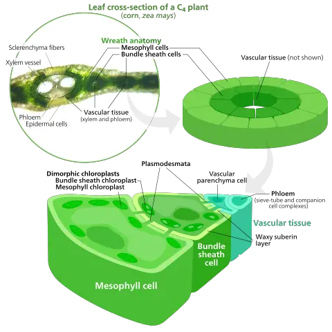
* Whereas the mesophyll tissue is composed of two layers of cells (spongy and palisade cells), the mesophyll tissue in monocots is largely composed of isodiametric cells (cells that appear spherical or polyhedral in shape).
Origin of Mesophyll Cells
Essentially, mesophyll cells make up the internal mesophyll tissue of a leaf. Here, these cells make up the cortex largely composed of parenchyma cells.
In vascular plants, the mesophyll layer, being a ground tissue, is the product of a group of cells known as ground meristematic cells which are themselves produced by cells of the apical meristem.
In plants, a group of cells located in the meristem (meristematic tissue) act as stem cells found in animals. As such, they divide to give rise to cells that differentiate to perform various functions in plants.
When cells of the ground meristem divide and differentiate, they may be distinguished into a number of tissues including the cortex, pith and pith rays. In the leaves, they give rise to the parenchyma cells of the mesophyll layer (palisade and spongy mesophyll cells) that are involved in photosynthesis.
This may be represented as follows:
Location
In order to clearly understand the location and arrangement of mesophyll cells, it's important to look at the general structure of a leaf.
A leaf is made up of a number of tissues that include the epidermis, the mesophyll layer, and the vascular tissue.
The epidermis composed of epidermal cells is the outer most layer that covers the upper (adaxial) and lower (abaxial) surface of the leaf. While the epidermis is a separate tissue from the other two, it acts as a protective layer that regulates material that enter or leave the cell.
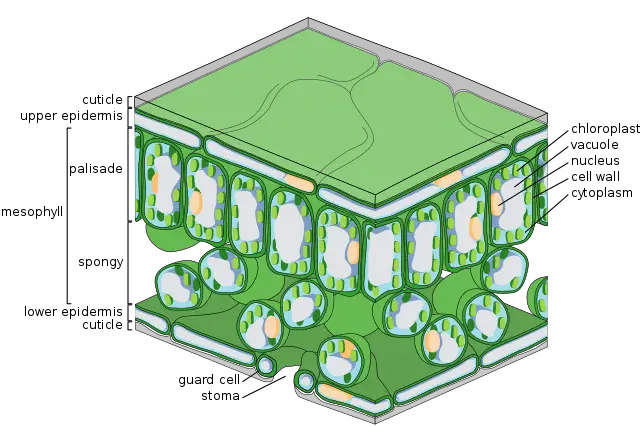
The mesophyll (ground tissue) is located between the upper and lower epidermis. Here, and particularly in dicots, the mesophyll is composed of two types of cells that include the palisade parenchyma cells located just below the epidermis and the spongy parenchyma cells that are located below the palisade cells and above the lower epidermis.
The vascular tissue, on the other hand, is located in the mesophyll layer where they are involved in the movement of material to the cells. The mesophyll layer, consisting of mesophyll cells, is therefore sandwiched between the upper and lower epidermis with the vascular bundles (xylem and phloem) running between its cells.
While the two types of cells form the mesophyll layer, they vary in morphology and serve different functions.
Structure
As already mentioned, the mesophyll layer is composed of two types of cells.
These include:
Palisade Cells
Palisade cells are part of the cells that collectively make up the mesophyll tissue in plant leaves. This layer (palisade layer) is located beneath the upper epidermis and is composed of cells that are columnar/cylindrical in shape.
In addition to a nucleus, some of the other important organelles of palisade cells include a cell membrane, a large vacuole, chloroplasts as well as a cell membrane among a few others. Between the cells (palisade cells are generally arranged in a vertical manner to each other beneath the epidermis) are slight separations that allow various materials to flow.
The structure and arrangement of palisade cells in the mesophyll tissue plays a crucial role in photosynthesis. Because of their shape (elongated and cylindrical) palisade cells contain many chloroplasts Palisade cells contain 70 percent of all chloroplasts. This is not only made possible by the shape of the cells, but also by the fact that compared to the other mesophyll cells, palisade cells are arranged in close proximity to each other.
In addition to these features, palisade cells are also well positioned to absorb more light required for photosynthesis. As already mentioned, palisade cells are located beneath the epidermis, which is itself a thin layer of cells. This allows palisade cells to absorb as much as is needed for the process of photosynthesis.
The structure/morphology of palisade cells is also beneficial for chloroplasts, and thus to photosynthesis is a number of ways.
These include:
Chloroplast movement - Light conditions have been shown to induce the movement of chloroplasts in a cell. In conditions where the amount of light available is low (low light conditions) chloroplasts move to and accumulate along the cell wall so that they are perpendicular to the incident rays.
When the amount of light is too high, they have been shown to move to regions of the cell so that they are not overly exposed. The capacity of chloroplasts to move (moved by specific structural proteins) within the cell is made possible by the fact that the elongated shape of palisade cells provide sufficient room for them to move and adjust their position with changes in light intensity.
Large vacuole - Although the shape of palisade cells allows them to move when need be, the large vacuole located at the central part of the cell restricts chloroplasts to the area along the cell membrane. This ensures that light easily reaches the chloroplasts for photosynthesis to take place.
Spongy Mesophyll Cells
Cells of the spongy mesophyll tissue are located below the palisade tissue and above the lower epidermis. Compared to the cells of the palisade layer, those of the spongy layer are spherical in shape or may be irregularly shaped (isodiametric) in some plants. These cells are also loosely packed which leaves a lot of spaces between the cells.
When viewed under the microscope, there are between 4 and 6 layers of spongy mesophyll cells that lie below the palisade cells. Like palisade cells, spongy mesophyll cells also contain such organelles as a nucleus, a vacuole, a cell membrane as well as chloroplasts among a few others.
The number of chloroplasts in these cells, however, is less compared to the number of chloroplasts found in palisade cells. Apart from the usual organelles in these cells, some of the spongy cells in leaves have also been shown to contain crystal inclusions in their vacuoles.
Whether in palisade or spongy cells, mesophyll cells that contain crystal inclusion (e.g. druse crystals) are shorter/smaller compared to the other cells in this region of the leaf.
* The thickness of the spongy parenchyma is between 1.5 and 2 times that of palisade tissue.
Depending on the type of plant, there are three variations of the spongy parenchyma:
- Typical spongy parenchyma cells
- Palisade-like spongy cells
- Aerenchymatous spongy cells
Although spongy mesophyll cells do not contain as many chloroplasts as those found in palisade cells, the nature of their arrangement plays an important role in photosynthesis. This is because being loosely packed enhances gas exchange during photosynthesis.
Function: Mechanism in Photosynthesis
Palisade Cells
Palisade cells are a type of parenchyma cells that contain most of the chloroplasts in plant leaves. Given that they are located beneath the upper epidermis, palisade cells are well positioned to absorb light required for photosynthesis.
In addition, their location ensures that carbon dioxide required for photosynthesis does not have to travel a long distance to reach the chloroplast.
As well, being located below the upper epidermis, which allows light, water, and gases to reach the cells easily, there are narrow spaces between the cells that ensure a large surface area of contact between the entire cell and air.
* The thin cell wall of palisade cells also allows gases to diffuse through with ease.
Because of the conditions provided by palisade cells, chloroplasts, located within these cells, are able to easily access the essential material required for photosynthesis to take place.
Here, the photosynthetic pigment known as chlorophyll in chloroplast absorbs given wavelengths of light which in turn provide the energy required for the photosynthetic reaction where carbon dioxide and water are used to produce a sugar molecule and oxygen.
Spongy Cells
Like palisade cells, spongy cells also contain some chloroplasts. Therefore, some level of photosynthesis also takes place in these cells. Unlike palisade cells, however, spongy cells are located deeper in the leaf below the upper epidermis and the palisade tissue.
With regards to photosynthesis, this is a disadvantage given that light does not penetrate to this region easily. As a result, spongy cells do not receive enough sunlight required for photosynthesis to occur ideally.
Although spongy cells are not well suited for photosynthesis processes, their arrangement are ideal for gaseous exchange. As previously mentioned, spongy cells are loosely packed above the lower epidermis. This creates large spaces between the cells which is ideal for gaseous exchange.
Small openings located on the epidermis allow such gases as carbon dioxide to enter the leaf and reach the mesophyll cells. On the other hand, photosynthetic processes in the mesophyll result in the production of oxygen.
The loosely packed cells (spongy cells) in this region of the cell allow these gases to be exchanged where oxygen is released while carbon dioxide is used for photosynthesis.
* In spongy cells, photosynthesis occurs at high light intensities.
Microscopy
Using an electron microscope, it's possible to not only clearly observe mesophyll cells, but also the architecture of the thylakoid membrane. However, for the purposes of observing mesophyll cells, a light microscope is sufficient.
Requirements
- Cassava cork
- Microscope - compound microscope
- Alcohol -30 percent, 50 percent, 70 percent, and 96 percent
- Safranin-O
- Clamp-on hand sliding microtome
- Young leaf
- Microscope glass slide and cover slips
- Preservation liquid (consisting of 70 percent alcohol and glycerin)
Procedure
With various samples, a vibratome is used for cutting in order to obtain thin sections that can be viewed under the microscope. However, with some samples, such as very thin leaves, alternative approaches may be used to cut in order to obtain the thinnest needed.
· For thin leaves, one of the methods suggested involves using cassava corks to hold and thus cut the sample. Here, a young, thin cassava stem is first cleaned and dried (under the sun or in the oven). A small leaf sample (1cm sq) is then cut out and inserted between the sliced cassava cork so that the sample is held between the sliced cork - Here, it's important to ensure that the cassava cork can fit in the hole of the mini microtome.
· Insert the cassava cork (with the sample held in between the sliced part) into the hole of the microtome and using the blade, cut the cork in order to obtain several transversal slices - Try to obtain very thin slices (almost transparent).
· Using a clean needle of the tip of a paint brush, carefully collect the section - the fresh section may be attached to the blade.
· Using a graded series of alcohol, dehydrate the sections obtained - 30 percent, 50 percent, 70 percent and 96 percent alcohol each for about half a minute.
· Remove the sample from the alcohol and ensure that all the alcohol has drained off.
· Place the sample in a mixture of 70 percent alcohol and glycerin (this is a preservation liquid) - This mixture may also contain such stains as 1 percent Safranin-O.
· Place the slide on a clean glass slide and cover using a clean cover slip - In the event that the sample is dry, a few drops of the preservation liquid may be added to prevent dehydration.
Observation
When viewed under the microscope, well prepared slices will display preserved mesophyll cells. Here, the epidermis will appear thin and darker while spongy cells will appear scattered below well organized palisade cells.
Return to Plant Biology overview
Return to Leaf Structure under the Microscope
See also info on Meristem cells of plants and Transgenic Plants
Return to learning about Guard Cells
Return to Organelles - Animal and Plant
Return from Mesophyll Cells page to MicroscopeMaster home
References
David S. Shatelet, et al. (2013). The Evolution of Photosynthetic Anatomy in Viburnum (Adoxaceae). Chicago Journals.
D. Metusala. (2017). An alternative simple method for preparing and preserving cross-section of leaves and roots in herbaceous plants: Case study in Orchidaceae.
Eiji Gotoh, et al. (2017). Palisade cell shape afects the lightinduced chloroplast movements and leaf photosynthesis. Scientific Reports.
J.V. van Greuning, P.J. Robbertse and N. Grobbelaar. (1984). The taxonomic value of leaf anatomy in the genus Ficus.
Keith Roberts. (2008). Handbook of Plant Science, Volume 1.
Nobuo Chonan. (1978). A Comparative Anatomy of Mesophyll Among the Leaves of Gramineous Crops. Faculty of Agriculture, Ibaraki University.
Links
https://mmegias.webs.uvigo.es/02-english/1-vegetal/v-imagenes-grandes/parenquima_clorofilico.php
https://worldwidescience.org/topicpages/s/spongy+mesophyll+cells.html
Find out how to advertise on MicroscopeMaster!


