What are the Functions of Lipids, Proteins, and Lipopolysaccharides on the Cell Membrane?
The cell membrane is an important barrier that separates the internal environment of a cell from the external environment. It separates the internal cell environment from the extracellular matrix in multicellular animals.
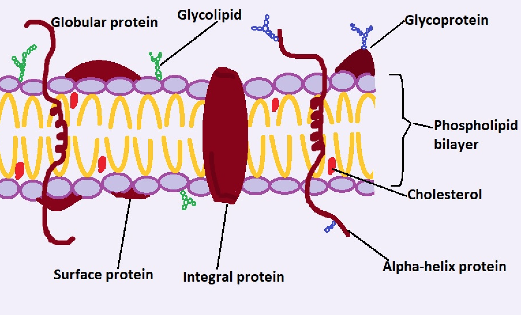
While it's commonly referred to as a phospholipid bilayer structure, the cell membrane also consists of several other components including proteins and carbohydrate groups.
Functions of Lipids in the Cell Membrane
Lipids are some of the most important components of the cell membrane, making up most of the structure. In general, the cell membrane has been shown to make up 50 percent of the membrane (by weight).
Here, however, it's worth noting that the structure consists of three main types of lipids.
These include:
Phospholipids (also known as phosphatides)
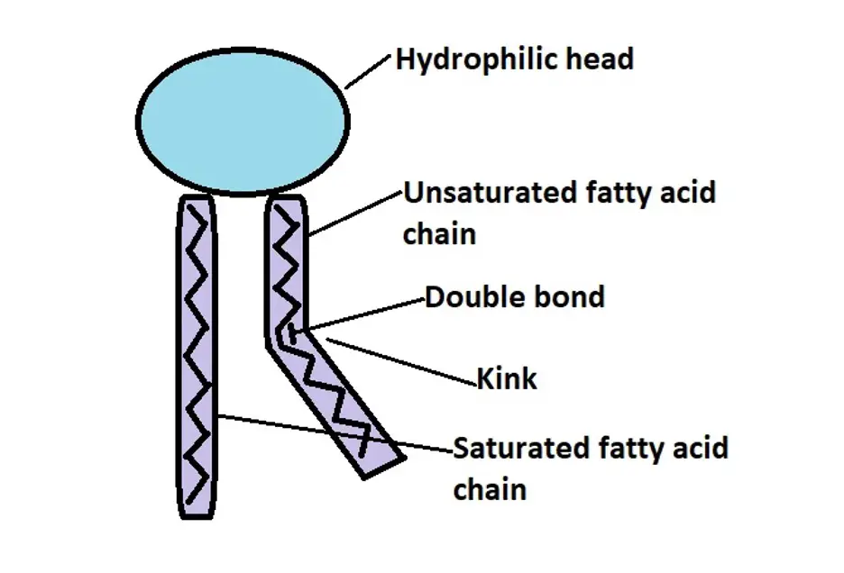
Phospholipids are the most abundant lipids of the cell membrane. As the name suggests, phospholipids consist of a phosphate group and two fatty acid chains (saturated and unsaturated fatty acid chains/fatty acyl tails).
Like triglycerides, they also have a glycerol to which the fatty acid tails and the phosphate group are attached (compared to triglycerides, phospholipids only have two fatty acid chains and a phosphate molecule).
While the phosphate group and glycerol molecule make up the head region of the phospholipid, the fatty acid chains are commonly referred to as the tails.
* The double bond in the unsaturated fatty acid chain caused the chain to bend (with a kink). This causes the chain to push the adjacent phospholipid thus creating a little distance between phospholipid molecules. In doing so, the kinks contribute to the overall fluidity of the membrane.
* In water, the phosphate group has a negative charge (because it donated hydrogen ions) which causes the phospholipid head to become hydrophilic.
Compared to the phosphate group, the fatty acids of the molecule do not have any charge and are also non-polar. For this reason, they are hydrophobic and thus repel water molecules.
Functions
- They form the bilayer of the cell membrane
The amphipathic nature of the phospholipids (having hydrophobic and hydrophilic regions) contributes to the main function of these molecules; formation of the phospholipid bilayer of the cell membrane.
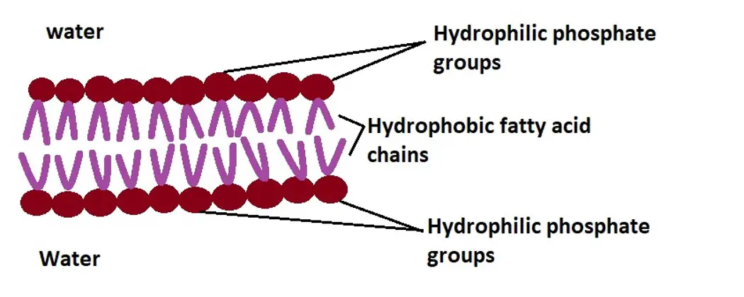
As mentioned, the phosphate group of the molecule is hydrophilic and therefore attracts water molecules. As well, the fatty acid tails which are hydrophobic in nature repel water molecules and will therefore face away from the water.
Due to this behavior, phospholipids form a layer where the hydrophilic groups face the water molecules while the hydrophobic tails face away from the water. This results in a phospholipid bilayer structure where the hydrophobic tails of each layer are localized in a hydrophobic region while the phosphate groups of each layer form the inner and outer surfaces of the structure.
The phospholipid is a crucial part of the membrane because it makes up the main barrier between the inner and outer regions of the cell.
Because the hydrophobic tails are never exposed to the aqueous environment (which contains water among other substances), the structure is kept stable. This is especially important given that it helps maintain the general structure/shape of the cell.
- Regulate the movement of substances in and out of the cell
The other important function of phospholipids is that they help regulate the movement of substances in and out of the cell. As already mentioned, phospholipids build the phospholipid bilayer which is the main barrier between the inner and outer environment of the cell.
As such, they also help control the type of substances that move in or out of the cell. In particular, this membrane plays an important role in the diffusion of small molecules (e.g., gases like carbon dioxide and small uncharged molecules) in an out of the cell.
Because they are very small, hydrophobic (e.g., benzene), or uncharged (e.g., ethanol, etc.), these molecules easily diffuse through the hydrophobic region and move into the cell or out of the cell depending on the concentration gradient. In the process, the phospholipids also play an important role in maintaining homeostasis.
* Because of the phospholipid bilayer which is made up of the phospholipids, the membrane is partially permeable and thus capable of allowing movement of some substances in or out of the cell.
- They act as electrical insulators
Because of the nature of the phospholipids that make up the phospholipid bilayer, charged ions cannot pass through the layer. They are dissolved in water and can therefore not easily diffuse through the hydrophobic region.
For this reason, the layer acts as an electrical insulator, keeping the right charge inside and outside the membrane. In the neurons, in particular, this function of the lipids helps propagate the proper speed of signals in the form of ions along the axons.
* While the inner and outer membrane contains phospholipids, the type of phospholipids they contain vary. Whereas the outer membrane contains phosphatidylcholine and sphingomyelin, the inner membrane consists of phosphatidylethanolamine and phosphatidylserine. - These are different classes of phospholipids.
* The distribution of different types of phospholipids vary between different types of cells.
Cholesterol
Cholesterol, a type of sterol, is also a membrane lipid that makes up between 25 and 30 percent of the total lipids in the cell membrane.
Like the phospholipid molecules, a cholesterol molecule also consists of a hydrophilic region (a polar head) and a hydrophobic chain (alkyl chain).
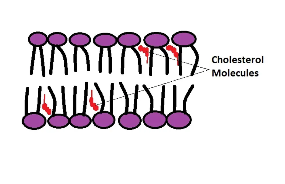
In the cell membrane, these molecules are located between the gaps created by fatty acid chains (of phospholipids). Here, the hydrophilic region of the cholesterol molecule is oriented towards the hydrophilic part of phospholipids while the tail of the cholesterol resides in the hydrophobic region of the membrane.
In the cell membrane, cholesterol has a number of important functions that include:
- Regulates movement of substances in and out of the cell
By occupying the spaces between the phospholipid molecules, cholesterol molecules decrease the permeability of the membrane by increasing lipid packing. This makes the membrane less permeable to a variety of smaller substances that would otherwise easily diffuse between the phospholipid molecules.
- Increases flexibility of the membrane
In the cell membrane, cholesterol has also been shown to contribute to the flexibility of the structure. In the absence of cholesterol, studies have shown that the phospholipid molecules can get much closer together, especially in the cold, which significantly reduces its flexibility. This can affect the fluidity of the cell membrane and the ability of some substances to pass through.
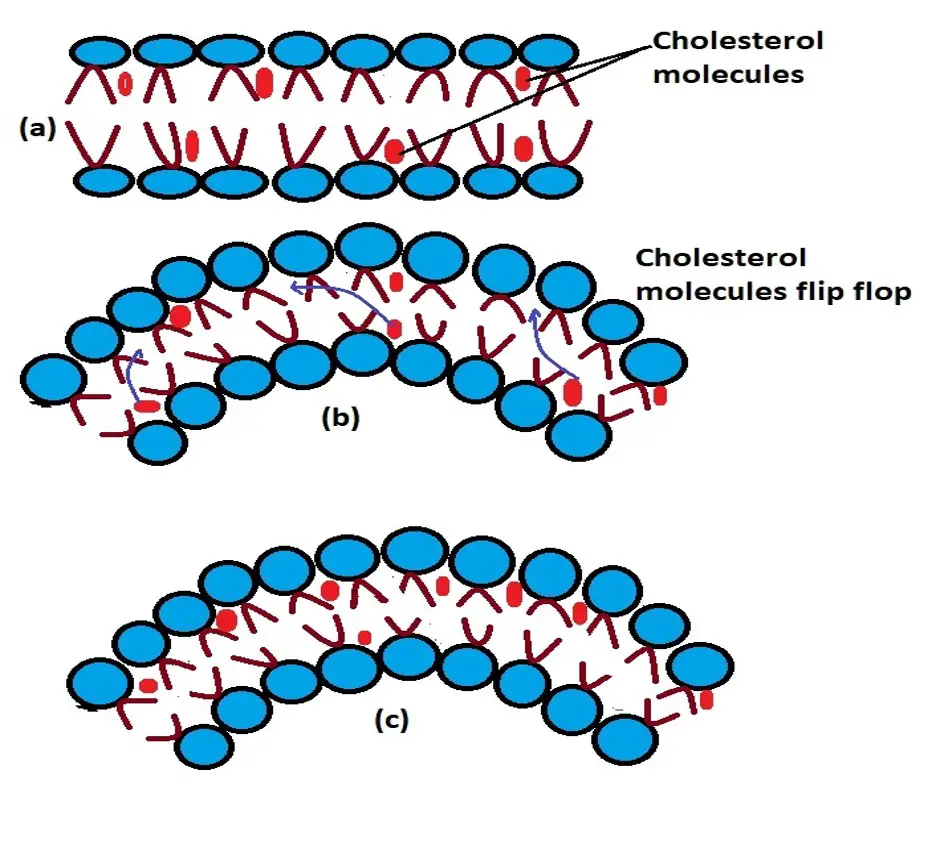
Based on research studies, cholesterol molecules in the membrane have been shown to change position and move to a given region through a process known as a flip-flop. This allows them to contribute to the flexibility of cells especially when they change shape.
A good example of this is when neutrophils squeeze between cells of the blood vessels (capillaries) in order to leave circulation and reach an infected site.
(a) Cholesterol molecules are evenly distributed in the membrane
(b) The membrane changes change and cholesterol molecules start to move (flip-flop)
(c) Cholesterol molecules have moved and are highly concentrated in one side of the structure where they enhance flexibility
- Enhance the stability of the cell membrane
In addition to promoting flexibility, cholesterol has also been shown to stabilize the cell membrane with changes in temperature. At high temperatures, the presence of cholesterol in the membrane increases the melting point of the membrane which in turn prevents the membrane from becoming overly permeable.
As well, intercalation of these molecules (cholesterol) between the phospholipids during low temperatures prevents the membrane from becoming very stiff which would otherwise affect how the cell functions. Therefore, the presence of cholesterol plays an important role in promoting the stability of the membrane.
Glycolipids
As the name suggests, glycolipids are the type of lipids that contain carbohydrate molecules. Here, a monosaccharide or oligosaccharide molecule is bound to the lipid through covalent bonds.
Compared to the other lipids, glycolipids are only found in the outer membrane (non-cytosolic side of the membrane). They can be found on the cell membrane of all eukaryotes and make up about 5 percent of the total lipids in the membrane.
* Like the other lipids, glycolipids are also amphiphilic molecules. As such, they consist of a hydrophilic region (the sugar group) as well as a hydrophobic region (the lipid moiety which is attached to the cell membrane)
Functions
On the cell membrane, glycolipids have a number of functions that include:
They act as surface receptors - Glycolipids on the surface of multicellular eukaryotic hosts act as receptors for various pathogenic bacteria. Here, the pathogen (or toxins) attaches to the host cell by binding to the receptor which in turn allows them to gradually invade the cell.
Signal transducers - Also known as cell signaling, signal transduction refers to the process through which molecular signals are transmitted from the exterior of a cell to the interior part of the cell.
During signaling, Glycosphingolipid, which is a type of glycolipid, associates with like molecules GPI-anchored proteins and growth factor receptors. A signal is then transmitted allowing the cell to respond effectively. This has also been shown to play an important role in cell proliferation.
Biosurfactant and in homeostasis - Essentially, biosurfactants are compounds with the capacity to reduce surface tension and interfacial tensions between surface molecules.
Because of their amphiphilic nature, glycolipids (e.g., Mannosylethritol lipids), produced by microorganisms like Candida antarctica act as biosurfactants.
Here, they have been shown to significantly reduce the antimicrobial activity of some of the agents used against them. As well, glycolipids like gangliosides which have a high affinity for calcium play an important role in synaptic transmission.
Functions of Proteins in the Cell Membrane
Like lipids, proteins also make up about 50 percent of the cell membrane (by weight). These proteins also make up about a third of the total proteins in the human body.
Membrane proteins are divided into two main categories that include:
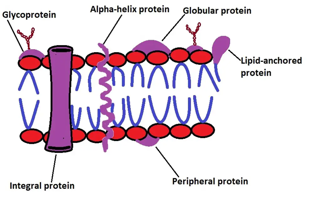
Integral proteins (intrinsic proteins) - These are permanent membrane proteins and include monotopic integral proteins, only attached to one layer/side of the phospholipid bilayer, transmembrane proteins (bitopic or polytopic) as well as some of the proteins associated with lipids (lipid-anchored proteins which are covalently bound to lipids e.g., Glycosylphosphatidylinositol).
Peripheral proteins (extrinsic proteins) - Peripheral membrane proteins, on the other hand, include surface proteins which are transiently associated with the membrane of the integral proteins of the membrane.
* Transmembrane proteins are also amphipathic (amphiphilic). As such, they have hydrophilic regions, located in the hydrophilic regions of the phospholipid bilayer, and hydrophobic regions which reside in the hydrophobic region of the bilayer - This is why these proteins are permanent residents of the cell membrane.
Transportation
One of the main functions of membrane proteins is the transportation of substances in and out of the cell.
In particular, these proteins are involved in two main types of transport that include:
Facilitated diffusion - Facilitated diffusion is a type of transport through which substances are transported down the concentration gradient (from where they are highly concentrated to where they are less concentrated).
The three types of proteins involved in facilitated diffusion are gated channel proteins, carrier proteins, and channel proteins.
This type of transport serves to move different types of substances including ions, amino acids, carbohydrates, and glycosylamines among others. These substances may be charged, too large, or polar and thus incapable of diffusing through the phospholipid bilayer.
While carrier transport proteins can actively transport molecules against their concentration gradient using ATP energy, facilitated transport does not need energy given that substances are often moving down their concentration gradient.
Active transport - Unlike facilitated diffusion, active transport involves the movement of substances against their concentration gradient across the cell membrane. Generally, active transport is divided into two main categories that include primary and secondary transport.
See also: Primary Diffusion Vs Active Transport
Primary Active Transport
Primary active transport is the type of active transport where energy in the form of ATP is used directly.
There are several types of primary active transport that include:
Uniporter (e.g., glucose transporter) - Only move a given type of substances in a single direction
Symporters (sodium iodide symporter) - Only move two substances in one direction
Antiporters (e.g., sodium and potassium pump) - Capable of moving two substances in opposite directions
Secondary Active Transport
The second type of active transport is known as secondary active transport. Unlike primary active transportation, this type of active transport relies on energy stored in the electrochemical gradient of ions to transport other substances against their concentration gradient.
Here, one of the best examples of this type of transportation is the transport of glucose through the symporter involved in the movement of sodium ions down the electrochemical gradient.
In this case, the energy produced as the ions are transported down the electrochemical gradient makes it possible for glucose to be transported through the symporter.
Cell Junctions
Cell junctions are structures (consisting of proteins) that connect cells to each other (cell-to-cell contact) or connect cells to the extracellular matrix.
These junctions are divided into three main categories that include:
Occluding junctions (tight junctions) - Tight junctions are protein complexes commonly found in epithelial tissue. Here, they connect cells together in a manner that prevents small molecules on one side of the epithelial sheet from leaking to the other side. They have been shown to contain more than 40 types of proteins (both cytoplasmic and transmembrane proteins).
Here, some of the transmembrane proteins that make up these junctions include claudins, occludin, and JAM proteins (junction adhesion molecule proteins). While tight junctions play an important role in creating a barrier between tissue spaces, they also control the movement of substances across the epithelium.
Communicating junctions (gap junctions) - As the name suggests, gap junctions are involved in communication, through the diffusion of ions and some molecules, between neighboring cells.
They primarily consist of integral membrane proteins known as connexins (connexin-associated proteins). Cell-to-cell communication established by gap junctions is crucial for a variety of activities including metabolic exchange in tissues as well as cell proliferation.
Anchoring junctions - Anchoring junctions are junctions that link neighboring cells as well as cells to the extracellular matrix.
These junctions have been divided into four main groups that include:
Adherens junctions - Involved in a number of functions including stabilizing adhesion between cells, regulating actin cytoskeleton, as well as regulating the signaling process between cells: These junctions are primarily composed of transmembrane proteins known as cadherins
Desmosomes - Desmosomes link neighboring cells and play a role in promoting the mechanical integrity of various tissues. By supporting the connection between cells in tissues, they have also been shown to promote morphogenesis.
Hemidesmosomes - Hemidesmosomes are similar to desmosomes. However, they are involved in connecting the basal surface of epithelial cells.
Cell-matrix adhesion complexes - These complexes connect cells to the extracellular matrix. They are primarily made up of integrins and adaptor proteins which provide structural support to the cells connected to the extracellular matrix.
The other important functions of membrane proteins include:
They act as markers - Some of the proteins in the cell membrane act as markers (allow for cell recognition). These proteins allow for cell recognition (e.g., recognition of specific proteins by antibodies)
Signal transduction - Some of the proteins are involved in cell signaling which allows the cell to respond
As enzymes - A number of proteins (e.g., Aminopeptidases) are located in the membrane where they are involved in various metabolic pathways
Functions of Lipopolysaccharides (endotoxins) in the Cell Membrane
Found in the outer membrane of Gram-negative bacteria, lipopolysaccharides are large glycolipids that consist of lipids covalently attached to long chains of sugar moieties known as oligosaccharides.
Overall, these large molecules (lipopolysaccharides) consist of three main parts which include lipid A which is the hydrophobic part of the molecule which is attached to the outer layer of the outer membrane, the core oligosaccharide (hydrophilic region connected to Lipid A), and the O antigen which is a repetitive glycan polymer.
* Some Gram-negative bacteria do not have lipopolysaccharides.
Function
Protection - One of the functions of lipopolysaccharides is that they help protect the bacterial cell from substances like bile salts and some antibiotics (lipophilic antibiotics).
Here, studies have suggested that in some Gram-negative bacteria, lipopolysaccharides are modified allowing the organisms to evade the actions of bile salts and some antibiotics used against them.
Pathogenicity - Following an infection, lipopolysaccharides (and specifically Lipid A) has been shown to activate the immune system. The toxic effect of the molecule is characterized by fever and even septic shock in some cases. In severe cases, this may result in organ failure and even death.
Stability - By increasing the negative charge of the membrane, lipopolysaccharides have been suggested to contribute to the stability of the membrane.
Return to learning about the Cell Membrane
Return to Different Cell Organelles and their Functions
References
Alberts, B. et al. (2002). Molecular Biology of the Cell. 4th edition.
Cooper GM. (2000). The Cell: A Molecular Approach. 2nd edition.
Renuka Malhotra. (2012). Membrane Glycolipids: Functional Heterogeneity: A Review.
Ronan Lordan, Alexandros Tsoupras, and Ioannis Zabetakis. (2017). Phospholipids of Animal and Marine Origin: Structure, Function, and Anti-Inflammatory Properties.
Links
https://xaktly.com/Lipids.html
Find out how to advertise on MicroscopeMaster!
