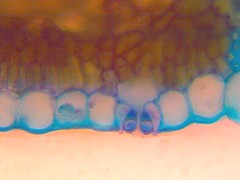
Guard Cells
Definition, Function, Structure of Stomata on Plants
Definition: What is a Guard Cell?
Essentially, guard cells are two bean-shaped cells that surround a stoma. As epidermal cells, they play an important role in gaseous exchange in and out of plant leaves by regulating the opening and closing of pores known as a stoma. In addition, they are the channels through which water is released from leaves to the environment.
As such, guard cells play a crucial role in photosynthesis by regulating the entry of materials necessary for the process. Apart from regulating gaseous exchange (as well as water release from leaves), they have also been shown to contain chloroplasts which also make them a site of photosynthesis.
Some of the factors that influence guard cell activities include:
- Humidity
- Temperature
- Light
- Carbon dioxide
- Potassium ions
- Hormones
* In Greek, the word "stoma" means mouth.
* Although stomata are commonly found in plant leaves, they can also be found in the stems.
Structure of the Guard Cells
As mentioned, guard cells are bean/kidney-shaped cells located on plant epidermis. As such, they, like trichomes and pavement cells, are also epidermal cells.
Between each pair of guard cells is a stoma (a pore) through which water and gases are exchanged. The opening and closing of these pores (collectively known as stomata) is made possible by the thickening and shrinking of guard cells on the epidermis.
* The number of stomata on a plant leaf/organ is highly dependent on the type of plant as well as its habitat.
Ultrastructure of Guard Cells
In different types of plants, guard cells have been shown to contain varying amounts of the typical cell organelles (among other structures) with some unique characteristics. For instance, as compared to the rest of a leaf, the cuticle of guard cells is more permeable to water vapor which in turn influences their activities/functions.
Guard cells have also been shown to have numerous ectodesmata. Here, the cuticle has also been shown to be more permeable to various polar substances. This is particularly important given that it is the concentration of these substances that influence the thickening and shrinkage of guard cells.
* On guard cells, the cuticle tends to be thicker on the outer parts.
* Cuticle permeability is also dependent on its chemical composition.
In young and developing guard cells, pectin and cellulose are gradually deposited into the plasmodesmata (a thin layer of cytoplasm). However, it disappears as guard cells mature while the few that are retained are devoid of any function.
There are also perforations on their walls that allow relatively large organelles to pass. For instance, plastids and mitochondria can pass through these perforations.
Various components can also be found in different types of guard cells in varying amounts and orientation.
In dumbbell-shaped guard cells, fibrils are radially in the outer wall. This orientation, however, may change with the thickening and shrinking of the cells. Apart from fibrils and microfibrils, a number of other substances have been identified in various guard cells.
In Zea mays, for instance, lignin has been identified in addition to cellulose. On the other hand, pectin has been identified in the guard cells of many plants.
Some of the organelles found in guard cells include:
· Microtubules - serve to orient cellulose microfibrils. They also contribute to the building and development of guard cells.
· Endoplasmic reticulum - The high amounts of rough endoplasmic reticulum present in guard cells are involved in protein synthesis. Apart from protein synthesis, ER is also involved in the formation of vacuoles and vesicles.
· Lysosomes - contain a number of molecules that contribute to the well functioning of the cell. These include; lipases, endopeptidases, phosphates, and DNAse.
· Lipid droplets - in guard cells are the intermediates in the synthesis of wax and cutin
· Nuclei - are centrally located in guard cells. They have been shown to change their general shape with shapes with the opening and closing of the stoma.
· Plastids - In guard cells, such plastids as chloroplasts vary in number from one plant to another. While some of these plastids may be poorly developed, others are well developed and capable of such functions as photosynthesis. In guard cells with functional chloroplasts, high amounts of starch during the night
· Mitochondria - High amounts of mitochondria can be found in guard cells (compared to mesophyll cells) which is evidence of high metabolic activities.
Stomata
Basically, stomata refers to both the pore (stoma) and the guard cells that surround them on the epidermis. Surrounding the guard cells are subsidiary cells that have been used to classify the different types of stomata.
While the stoma (pore/opening) is the channel through which gases enter the air spaces in leaves, opening, and closing of these openings is regulated by guard cells located on the epidermis.
Classification of Stomata
Generally, stomata are classified based on distribution and structure.
Types of stomata based on distribution/placement:
· Water lily type - are located on the upper epidermis of leaves. They can be found in many aquatic plants such as the water lily.
· Apple type (mulberry type) - are stomata that are typically found on the lower surface of leaves. As such, they can be found in such plants as walnut, apples, and peach among others.
· Potato type - A majority of these stomata can be found on the lower surface of leaves while a few may be found on the upper surface. As such, they are typically found in amphistomatic and anisostomatic leaves (e.g. potato, tomato, cabbage, etc.)
· Oat type - are found in isostomatic leaves (where stomata are distributed on the upper and lower surface of the leaves)
· Potamogeton type - are either absent or non-functional as is the case in submerged aquatic plants.
Based on Structure
· Anomocytic - A small number of subsidiary cells surround the stomata. For the most part, these cells (subsidiary cells) are identical to the other epidermal cells.
· Cruciferous - The stoma is surrounded by three types of subsidiary cells that vary in size.
· Paracytic - The stoma is surrounded by two cells (subsidiary) that are arranged in a parallel manner to the axis of the guard cells.
· Graminaceous - Here, the guard cells are dumbbell-shaped. With subsidiary cells arranged parallel to them.
· Diacytic - The stoma in this classification is two guard cells. The wall of the subsidiary cells surrounding the stoma is at a right angle to the guard cells.
· Cyclocytic - Here, a minimum of four subsidiary cells surround the guard cell.
* 80 to 90 percent of transpiration occurs through the stomata. Water is also lost through lenticular and cuticular transpiration.
* Only a small amount of water absorbed (about 2 percent) is used for photosynthesis in plants.
Adaptations
Guard cells have a number of adaptations that contribute to their functions.
These include:
They have perforations through which solutes and water enter or leave the cells - This is one of the most important adaptations of the guard cells because the movement of solutes and water in and out of guard cells cause them to shrink or swell. In turn, this results in the closing or opening of the stoma/pore through which water and gases are exchanged.
They contain chloroplasts - Although they do not contain as many chloroplasts as mesophyll cells, guard cells have been shown be the only epidermal cells with chloroplast.
As such, guard cells of soma plants are photosynthetic sites where sugars and energy are produced. It's worth noting that chloroplast is either absent or inactive in some guard cells.
They contain hormone receptors - allowing them to respond appropriately to changes in their environment. For instance, water scarcity in the soil causes the release of a hormone (abscisic acid (ABA)).
This hormone is transported from the root cells to the receptors on guard cells which in turn causes the guard cells to close the stoma in order to prevent excessive water loss.
Bean/kidney-shape - The shape of guard cells is convenient for the closing and opening of the stoma to regulate gaseous exchange and release of water.
Guard cells are surrounded by a thin, elastic outer wall - contributes to the movement of water and solutes in and out of the cell.
Location - Depending on the habitat, guard cells may be located on the upper or lower surface of the leaf. This regulates the amount of water lost to the environment.
In most aquatic plants, guard cells, and thus the stomata, are located on the upper surface of the leaf which allows for more water to be released into the environment. However, for plants in hotter/dry areas, these cells are located on the lower surface of the leaf and tend to be fewer in number.
Closing and Opening Mechanism
One of the most important functions of guard cells is to control the closing and opening of the stoma/pores. While the opening of these pores allows water to be released into the environment, it also allows carbon dioxide to enter the cell for photosynthesis (as well as the release of oxygen into the environment). For this reason, guard cells play a crucial role in photosynthesis.
Based on a number of studies, such factors as light intensity and hormones have been shown to influence the swelling or shrinkage of guard cells and thus the opening and closing of the pores.
Here, with regards to pore opening, these factors influence water uptake into the cell causing the guard cells to inflate. This inflation/swelling results in the opening of the pores which in turn allows for gaseous exchange (as well as the release of water/transpiration).
While the process sounds to be a simple one, the signaling pathway that influences guard cell activities is yet to be fully understood. For this reason, a number of theories have been presented (and refuted) to describe the entire process/mechanism. Regardless, several aspects are well understood and will be highlighted in this section.
Theories aimed at explaining the movement of water in and out of guard cells include:
· pH theory – An increase in the concentration of hydrogen ions causes a decrease in pH which in turn results in the conversion of glucose-1-phosphate to starch.
· Starch-sugar theory - Conversion of starch to sugar causes the osmotic potential to increase thus drawing water into the guard cells.
· Proton-potassium pump theory - Through a sequence of events, potassium ions are transported into the guard cells during the day increasing solute concentration and drawing water into the cell.
· Active K+ transport theory - An increase in potassium ions is caused by the conversion of starch to phosphoenolpyruvate and consequently malic acid.
Carbon Dioxide Sensing and Signaling
One of the factors that influence the swelling and shrinkage of guard cells is carbon dioxide concentration. In cases of high carbon dioxide concentration in the atmosphere, studies have shown anion channels to be activated causing potassium ions to move out of the cells. At the same time, chloride is released from the cells ultimately reusing in the depolarization of the membrane.
With solutes moving out of the cell, their concentration out of the cell increases as compared to that inside the cell. As a result, water is forced out of the cell through osmosis. In turn, this causes the cell to shrink and close the aperture/pore.
* Malate is suggested to be an intermediate effector between the gas (carbon dioxide) and activation of the channel.
* At low partial pressure of carbon dioxide in the atmosphere, the reverse occurs.
Abscisic Acid (ABA) Sensing and Signaling
In different types of plants, ABA (a plant hormone) has a number of functions ranging from controlling the germination of seeds to its impact on guard cells.
In such environmental conditions as drought or increased salinity in soil, roots have been shown to produce this hormone in higher amounts. The detection of this hormone by guard cells causes changes in the intake or removal of ions from the cells which in turn causes the opening or closing of the stoma. Here, a subunit of Mg-chelatase was shown to bind the hormone and thus serve as the intermediate.
In instances of high amounts of ABA, the efflux of anions as well as potassium through the channels occurs. At the same time, importation of potassium ions is inhibited which prevents the ions from moving into the cell (this would otherwise cause a high concentration of solutes in the cell).
With high solute concentration outside the cell, water is forced out through osmosis, which in turn reduces turgor pressure of the guard cells. In turn, this causes the aperture to close, preventing the cells to lose any more water.
* Under normal environmental conditions, stomata open during the day to allow for intake of carbon dioxide and close at night when light-independent reactions (photosynthetic reactions) take place.
* At night, water enters the subsidiary cells from the guard cells which causes them to become flaccid (reducing turgor pressure in guard cells) and thus causing stoma to be closed.
See also Mesophyll Cells and Meristem Cells.
Return to studying Leaf Structure under the Microscope
Return from Guard Cells to MicroscopeMaster home
References
Cecie Starr. (1991). Biology: Concepts and Applications.
June M. Kwak, Pascal Mäser, Julian I. Schroeder. (2009). The Clickable Guard Cell, Version II: Interactive Model of Guard Cell Signal Transduction Mechanisms and Pathways.
J. M. Whatley. (1971). The Untrastructure of Guard Cells of Phaseolus Vulgaris.
Mareike Jezek and Michael R. Blatt. (2017). The Membrane Transport System of the Guard Cell and Its Integration for Stomatal Dynamics.
Sallanon Huguette, Daniel Laffray, and Alain Coudret. (1993). Structure, ultrastructure and functioning of guard cells of in vitro rose plants. ResearchGate.
Links
https://www.ncbi.nlm.nih.gov/pmc/articles/PMC3258058/
https://www.cell.com/current-biology/pdf/S0960-9822(01)00358-X.pdf
Find out how to advertise on MicroscopeMaster!
![Confocal image of Arabidopsis stomate showing two guard cells by Alex Costa[CC BY 2.5(https://creativecommons.org/licenses/by/2.5)] Confocal image of Arabidopsis stomate showing two guard cells by Alex Costa[CC BY 2.5(https://creativecommons.org/licenses/by/2.5)]](https://www.microscopemaster.com/images/Plant_stoma_guard_cells.png)

![Overview on mechanisms & ion channels involved in turgor regulation of guard cells, controlling stomatal aperture in plants.By June Kwak,University of MarylandJune Kwak, Pascal Mäser[Public domain] Overview on mechanisms & ion channels involved in turgor regulation of guard cells, controlling stomatal aperture in plants.By June Kwak,University of MarylandJune Kwak, Pascal Mäser[Public domain]](https://www.microscopemaster.com/images/Guard_cells_mechanisms.png)


