Order Piroplasmida
Classification, Characteristics and Life Cycle
Definition: What is the Order Piroplasmida?
Order Piroplasmida, consisting of organisms known as piroplasms, is an Order ranked under the Phylum Apicomplexa. Like members of the Order Haemosporida, piroplasms parasitize erythrocytes (red blood cells) of mammals and birds. As such, they cause diseases in both human beings and animals with low pathogenicity being demonstrated in wildlife.
The group consists of three main genera namely, Babesia, Theileria, and Cytauxzoon that are widely distributed across the globe. Of these, Babesia and Theileria have been associated with significant economic losses due to their negative impact on domestic production animals.
* Depending on the species, asexual reproduction may occur in red cells or leukocytes.
Some of the species that belong to the Order Piroplasmida include:
- Theileria parva
- Theileria lestoquardi
- B. microti
- Bubesia equi
- Babesia gibsoni
Classification of the Order Piroplasmida
· Phylum: Apicomplexa - This is a large group that consists of parasitic alveolates (protists). Although they exhibit significant diversity with regards to morphology, they are all characterized by the presence of an apical complex structure.
· Class: Conoidasida - Created by Mehlhorn et al in 1980, this class consists of organisms characterized by a tip at one end of the organisms' outer membrane and vesicles (rhoptries and micronemes) that open at the anterior part of the cell
· Subclass: Coccidiasina - A group consisting of single-celled, spore-forming, obligate intracellular parasites of the class Conoidasida
· Order: Piroplasmida - Pear-shaped parasites (within red blood cells) divided into three major genera (Babesia, Theileria, and Cytauxzoon)
Characteristics of the Order Piroplasmida
General Morphology
As members of the Phylum Apicomplexa, Piroplasms share a number of morphological characteristics with each other. For instance, all Piroplasms have apical complex organelles including dense granules, micronemes, and rhoptries.
Within red blood cells, many of the parasites present a pear shape which is the basis of their name. However, there are also various morphological differences that have been used to describe the different development stages of the organisms and even divide the groups into several subgroups.
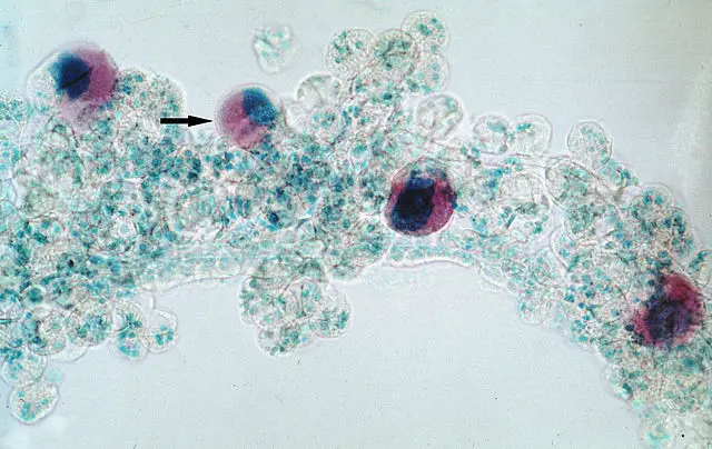
Based on morphology, the genus Babesia is divided into two main groups that include small Babesias (ranging between 1.0 to 2.5um in length e.g. B. bovis and B. microti) and large Babesias that range between 2.5 and 5.0 um in length (e.g. B. cabalii and B. bigemina). Although these parasites have a pear-shaped appearance in red cells, they may also appear spherical or amoeboid.
* The name piroplasm (derived from the word pyriform) is due to the pear-shape of these parasites within red blood cells of the host.
Distribution
Given that these parasites are spread by ticks, they can be found in different regions across the globe. However, the prevalence of different groups varies from one region to another. Whereas Babesiosis (caused by Babesia) is endemic across the northeastern and upper Midwestern parts of the United States, it is not as common in other Europe, South America, and Africa. East Coast Fever (theileriosis) which is caused by Theileria is particularly common in parts of Africa and has been reported in parts of America.
Distribution of these organisms is largely dependent on the genus as well as given environmental conditions (temperature, etc).
Based on various studies, the prevalence of these organisms has also been shown to be seasonal and thus dependent on the weather. In Bangladesh, weather conditions (between April and September (characterized by higher temperatures and humidity, etc) have been shown to influence the multiplication of ticks and consequently an increase of Theileria.
In other countries (Parts of the United States and Australia), these changes have been associated with increased infections and heavy economic losses due to infected production animals.
Reproduction and Life Cycle
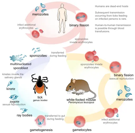
The life cycle and reproduction in piroplasms can be divided into the following categories depending on the type of organisms:
- Intra-leukocytic schizogony (by various Theileria species)
- Intra-erythrocytic merogony (post-intra-leucocytic schizogony phase in Theileria species)
- Intra-erythrocytic merogony by Babesia species
- Gamogony - sexual reproduction
- Sporogy- involves the formation of sporonts which in turn forms the sporoblasts
Schizogony
Schizogony refers to the asexual mode of reproduction common among Theileria sensu stricto, Theileria equi and Cytauxzoon spp. Unlike Merogony, this mode of reproduction involves the invasion of leukocytes by sporozoites to produce merozoites. Before the sporozoites invade these nucleated cells (leukocytes) studies have shown them to alter their metabolism.
Following a tick bite, which introduces the sporozoites into the body of the host, the parasites are suggested to come into contact with the cell membrane of the host cells randomly. Following this contact, they attach and penetrate the cell without the use of the apical structure. Rather, apical organelles excrete proteins that allow the organism to establish itself in the cytoplasm of the host cell.
As the parasite invades these nucleated cells, it has also been shown to shed its own coat.
The whole process takes about three minutes and involves the following stages:
· Recognition and attachment - recognize and attach to the nucleated blood cells
· Junction formation - a junction is formed between the parasite and the cell membrane of the target cell
· Zippering - internalization of the parasite
· Separation and dissolution - The parasite enters the cell as the cell membrane of the host cell closes
* Unlike other intracellular parasites, internalization of these parasites does not result in the formation of parasitophorous vacuoles.
Within these cells, the sporozoites undergo changes to form multinucleated schizonts that in turn produce uninucleated merozoites which enter circulation and invade red blood cells.
* Internalization of Theileria sporozoites into leukocytes result in cell damage.
Merogony
This mode of asexual reproduction is common among Babesia parasites and involves intra-erythrocytic multiplication. As is the case with Theileria sporozoites, the first contact between Basesia parasites and red cells of the host occurs through random collisions. However, internalization of these invasive forms of the parasite into the cells involves the use of apical structures.
Here, these structures aid in forming the junction between the cell membrane of the host and the organism. Once the junction has been established, proteins secreted by secretory organelles of the apical mediate parasite penetration into the cell.
Apart from penetration, these proteins also influence orientation of the parasite within the red cells of the host. The whole process takes a few minutes and does not result in the formation of parasitophorous vacuole.
Within the cells, these sporozoites develop to form trophozoites that undergo asexual division to form merozoites (this is known as merogony). This multiplication process causes the cell to rupture and release numerous merozoites which then infect new red blood cells.
* Depending on the species, merozoites may have a piriform or ovoid in shape and may occur in pairs or tetrads. Over time, however, they have been shown to become polymorphic with invaginations that result in pseudopods.
Gamogony
Gamogogy is a sexual mode of reproduction and normally occurs in the gut of the vector host (tick). Sexual stages of piroplasms are known as gametocytes and the first stages of these forms are found in the red blood cells of mammals.
Following a blood meal, these forms are ingested by ticks (vector host) and migrate to the lumen of its gut. Although they do not reproduce, they differentiate to form gametes in this location.
* Based on morphological studies, these gametocytes have been shown to be relatively larger in size with an unusual shape.
* Unlike the asexual forms, gametocytes are not destroyed in the gut lumen of the tick.
Once they are ingested along with red blood cells, gametocytes start to re-organize the cytoplasm and undergo development when the red cells are lysed. Here, the metamorphosis of these gametocytes produces gametes known as spiky-rayed stages or Strahlenkörper. The gametes then undergo division to form haploid gametes.
For some of the species (e.g. T. equi) microgametes and macrogametes can be distingushed from each other based on their general morphology. However, gametes do not differentiate into the two types of gametes in some of the species (e.g. in Babesia sensu stricto). Rather, they form two different gamete populations.
Following the formation of gametes, fertilization is stimulated by close contact between the gametes. This process involves the formation of filamentous structures between the membranes of gametes in contact with finger-like protrusions of opposite gametes penetrating the other. This process produces motile zygotes often referred to as ookinates.
Once the zygote is formed, it penetrates the peritrophic matrix located in the gut lumen of the vector host. Here, with the aid of enzymes released by an arrowhead structure of the zygote, the zygote invades epithelial cells of the gut where it develops further. Penetration into these cells allows the zygote to rest in the cytoplasm surrounded by cell organelles of the host cell.
Here, the zygote has been shown to undergo morphological changes and lose the arrowhead as it becomes spherical in shape. This is then followed by meiotic division to form kinetes that are released into the hemolymph of the tick. They are then covered by a fuzzy coat as they prepare for more division.
* Kinetes may invade various parts of the tick body and multiply asexually or invade the salivary gland where they undergo sporogony.
* In the salivary glands of the vector host, kinetes develop to form sporozoites that are transmitted to the secondary host (mammals) when the tick feeds.
Sporogony
Unlike the other processes mentioned above, sporogony is an asexual process that results in the formation of spores. In piroplasms, this starts when kinetes invade the salivary gland of the tick. Here, the kinetes begin to grow in size and develop into sporonts (polymorphous single-membrane syncytium). These sporonts then develop into sporoblasts. These sporonts remain dormant until the tick attaches to a host.
Piroplasms are responsible for a number of diseases in both animals and human beings.
Some of the diseases caused by these organisms include:
- East Coast Fever - Caused by T. parva
- Tropical theileriosis - Caused by T. annulata
- Oriental theileriosis - Caused by T. orientalis
- Babesioisis - caused by Babesia species
Return to Human Parasites under the Microscope
Return to learning about Parasitology main page
Return to learning about Protozoology main page
Return from Order Piroplasmida to MicroscopeMaster home
References
Larry Vogelnest and Timothy Portas. (2019). Current Therapy in Medicine of Australian Mammals.
Mary J. Homer, Irma Aguilar-Delfin, Sam R. Telford III, Peter J. Krause and David H. Persing. (2000). Babesiosis.
Marie Jalovecka, Ondrej Hajdusek, Daniel Sojka, Petr Kopacek and Laurence Malandrin. (2018). The Complexity of Piroplasms Life Cycles.
Ramgopal Laha. (2015). Morphology, epidemiology, and phylogeny of Babesia: An overview.
Links
https://instruction.cvhs.okstate.edu/kocan/vpar5333/5333iim.htm
Find out how to advertise on MicroscopeMaster!


