Protozoology
Definition, Examples, Classification and Characteristics
Introduction: What is Protozoology?
Essentially, protozoology is a branch of zoology that is concerned with the study of protozoa. As such, it may simply be described as the science of protozoa (microscopic eukaryotes that either exist as parasites or free-living organisms).
Here, protozoologists focus on the different types of protozoa, their respective characteristics, as well as how they interact with their surrounding (as parasites or free-living organisms).
As with any other organism in nature, protozoologists not only consider the pathogenic aspect of these organisms, but also some of the other important roles (in the food chain, degradation etc) they play as components of their respective ecosystems.
As a science of protozoa, protozoology is also related to the following fields:
- Microbiology
- Medical biology
- Immunology
- Zoology
- Parasitology
Classification
A number of approaches have been used to classify protozoa in protozoology. Whereas some protozoa exist as free-living organisms in various environments others exist and survive as parasites.
Classification of Protozoa Based On Locomotion
Based on the mode of locomotion, protozoa can be divided into 4 main groups.
These include:
Amoeboids (Sarcodina)
Amoeboid protozoa include protozoa that move by using temporary protoplasmic outgrowths known as pseudopods.
Some of the protozoa that belong to this category include:
- Radiolarians
- Entamoeba histolytica
- Pelomyxa
Flagellates (Mastigophora)
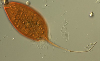
Protozoa classified as flagellates are, for the most part, single-celled organisms that use flagella for locomotion. Depending on the species, they may possess one or several structures at a given stage of their life cycle.
Some examples of flagellate protozoa include:
- Trypanosoma
- Leishmania
- Trichomonas
Ciliates (Phylum Ciliophora)
Unlike flagellates, ciliate protozoa use structures known as cilia for locomotion. Compared to flagella, cilia are shorter and tend to be present in large numbers. Paramecium and Blantidium species are some of the protozoa that use cilia for locomotion.
See more about Cilia and Flagella here.
Sporozoa
Unlike the other three, Sporozoa is a large phylum of parasitic protozoa that do not possess any structure for locomotion. As such, they may be described as non-motile protozoa. Malaria parasites that reside in red blood cells are good examples of Sporozoa.
Branches of Protozoology
Based on ecology/habitat, protozoa can be divided into several categories. For instance, whereas some protozoa can be found living freely in terrestrial environments (soil) others live as parasites in different animals.
As a result, protozoology as a field of study is divided into the following categories (branches):
Soil Protozoology
Established in the 1920s as a science by D.W. Cutler (an English Microbiologist), soil protozoology is the branch concerned with the study of soil protozoa. Currently, well over 1,600 species of protozoa have been identified in various terrestrial habitats with a similar number yet to be identified and described.
In terrestrial environments, protozoa move from one point to another by using such structures as cilia, flagella, and pseudopodia.
Based on locomotion, then, soil protozoa can be divided into the following groups:
Since the 1920s, specialists have continually added information into the field allowing it to be well established as it is today.
Characteristics of Soil Protozoa
A majority of soil protozoa are small in size, but several times larger than bacteria (ranging between 5 and 500um in diameter). The size is largely dependent on the type/species of protozoa.
Whereas flagellates range between 5 and 20um in diameter, ciliates are larger, ranging between 10 and 80um in diameter. However, they are still smaller when compared to Sarcodina (amoebas) some of which are large enough to be seen with the naked eye (e.g. Chaos carolinense which can grow to be 5mm in length).
Depending on the species, soil protozoa inhabit soil in great numbers, making up between 10,000 and 1 million individuals per gram of dry mass. Subject to environmental conditions, they are able to produce many generations annually which allows them to maintain the high numbers mentioned above.
* Their small size plays an important role in their survival. Because of their small size, soil protozoa are able to survive in very small films of moisture in their environment.
While given species are either omnivorous or tend to feed on certain fungi, a majority of soil protozoa have been shown to feed on bacteria for the most part. Through their feeding behavior, they have also been shown to play an important role in the flow of nutrients which in turn contributes to the proper growth of plants and other soil organisms such as earthworms.
* During unfavorable environmental conditions, soil protozoa produce protective cysts that allows the organism to survive harsh environmental conditions for a period ranging between several months to a few years.
* Some species of protozoa have also been shown to feed on other protozoa: Those smaller than themselves.
While soil protozoology is largely concerned with the study of soil-dwelling protozoa, some books will also touch on free-living protozoa found in aquatic environments.
This is because some of the marine and fresh-water protozoa also tend to contribute to various terrestrial ecosystems. While such protozoa as Dinoflagellates and Radiolaria are found in marine environments, their shells (after they die) make up deposits such as limestone.
Some species, however, have been shown to live in soil and aquatic environments. For instance, in New Zealand, five ciliate species are known to live in both freshwater and soil environments.
Examples of the protozoa found in soil include:
- Colpoda sp.
- Bodo sp.
- Naegleria species
- Paramecium
- Oikomonas termo
- Balantiophorus minutus
Medical Protozoology: Parasitic Protozoa
Unlike soil protozoology, medical protozoology is a branch that is largely concerned with the study of parasitic protozoa that infect and cause diseases in humans. Here, protozoologists focus on biology (life-cycle, morphology, etc) of infective/disease-causing protozoa as well as the clinical manifestations of the diseases they cause.
* According to studies, about a fifth of all protozoa exist as either parasites or symbionts with flagellates exhibiting the most diverse form of parasitism.
Some of the most important protozoan parasites in medical protozoology include:
- Giardia
- Cyclospora
- Cryptosporidium
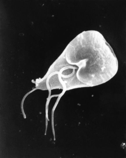
Giardia
Found in contaminated water, food or soil across the globe, Giardia is a single-celled parasite that causes giardiasis (diarrheal illnesses). Although the parasite can be spread in a number of ways (contaminated foods and fruits etc) water has been shown to be the most common mode.
* Since giardiasis is a result of consuming water/foods contaminated with feces from an infected person, the infection is particularly common in areas with poor sanitation.
Characteristics
The life cycle of Giardia species such as G. duodenalis consists of two main stages; the trophozoite stage and the non-proliferative cyst stage. Giardia cysts, which tend to be environmentally robust, are very persistent and thus capable of surviving various harsh environmental conditions.
In water, Giardia cysts have been shown to be highly resistant to various disinfectants used to treat water (e.g. chlorine). This not only allows them to survive for a long period of time outside the body, but also significantly contributes to their infective nature. When the cyst is consumed, along with infected water or foods, it transforms (through several asexual and sexual cycles) into a flagellated trophozoite that proliferates in the upper small intestine where it causes giardiasis.
* Giardiasis is more common in children given that they are more likely than adults, to consume contaminated water and foods/fruits
* In the small intestine, Giardia causes giardiasis (diarrhea) by attaching to the wall of the intestine and interfering with the absorption of fats and carbohydrates
Some of the other characteristics of Giardia include:
- Predominantly asexual
- Use adhesive discs to attach to the epithelial cells
- Characteristics of Guardia infections have been associated with affected enzyme activities in the gut
Cyclospora
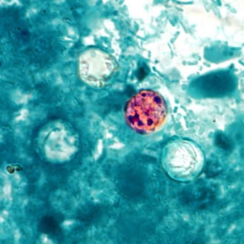
Cyclospora is a microscopic (single-celled) parasite that measures between 8 and 10 microns in diameter. Like Giardia, the parasite is spread by consuming foods or water contaminated by infected fecal matter containing the parasite. Therefore, it's also common in areas with poor sanitation.
* Although Cyclospora species (e.g. C. cayentanensis) were isolated as early as the 1870s, it is still considered an emerging pathogen.
* Cyclospora species make up a subclass of parasites referred to as Coccidia. These single-celled organisms are characterized by the fact that they form spores.
General Characteristics
Members of this genus (e.g. C. muris, C. felis, C. meleagridis, and C. baileyi) are taxonomically related to Toxoplasma and Cryptosporidium.
The life cycle of Cyclospora starts when the non-infectious oocytes are excreted into the environment along with feces. Several days/weeks after excretion, sporulation of the oocysts (spherical in shape and measure between 8-10um in diameter) begins under conducive environmental conditions (temperatures between 22 and 32 Degrees Celsius).
Here, the sporonts are divided into two sporocysts each of which consists of elongated sporozoites (measuring about 1.2 × 9 um), which are the infective agents. Once they are ingested (in contaminated water or foods), the sporozoites are released from the cyst and invade the epithelial cells of the jejunum where they obtain nutrition. It is also within these cells that they undergo various stages of both sexual and asexual reproduction to produce oocysts that allow the life cycle to continue.
* In humans, Cyclospora is responsible for an intestinal illness known as Cyclosporiasis. This is characterized by watery diarrhea.
* Little is known about how the parasite actually causes intestinal illness (pathogenic mechanism).
Some of the other characteristics of Cyclospora sp. include:
· Unsporulated oocysts are excreted about 60 days following the initial infection
· Parasite can be found throughout the world
· Majority of gametes (during reproduction) have been shown to form female gametes (macrogametes)
· In the environment (external to the host) Cyclospora form a cyst that allows them to survive harsh environmental conditions
Cryptosporidium
The genus Cryptosporidium falls under the phylum Apicomplexa and is composed of small coccidian protozoan parasites that tend to cause digestive and respiratory infections in vertebrates.
Because they lack distinct morphologic characteristics as well as strict host specificity, it proves rather challenging to describe new species of the genus for some time with only a few species being described in the early 2000s.
Some of these included:
- C. wrairi
- C. felis
- C. cannis
- C. meleagridis
- C. serpentis
While a majority of these species affect animals, a few have been shown to affect human beings and cause diseases.
Characteristics - Life Cycle and Infection
The life cycle of Cryptosporidium species starts when the oocysts are excreted through feces by an infected host. Unlike those of Cyclospora, the oocysts of Cryptosporidium are sporulated and contain 4 sporozoites.
Oocysts of Cryptosporidium may also be released into the environment through respiratory secretions given that the parasite has been shown to cause respiratory infections. Outside the body of the host, the oocysts are resistant to various harsh environmental conditions due to the presence of a two-layered oocyst wall that surrounds the sporozoites.
While the oocysts are immediately infectious once they are released into the environment (in water etc), they can survive for a long period of time outside the body due to the presence of the protective layer.
Once the oocysts are ingested, along with infected water, sporozoites excyst in the lumen of the intestine and penetrate the cells where they develop into trophozoites. Here, the trophozoites undergo asexual division to form merozoites some of which undergo sexual stages to form gametes that form the zygotes. It's these zygotes that then develop to oocysts that are released into the environment through feces allowing the life cycle to continue.
* Intestinal and respiratory illnesses are caused when the sporozoites parasitize the epithelial cells in these parts of the body. Infection of the small intestine causes diarrhea while infection of the respiratory system may result in a persistent cough.
Some of the other characteristics of Cryptosporidium sp. include:
- Complete their life cycle in a single host
- Cause acute infections in humans. For immunocompromised individuals, however, infections can be severe
- Outside the body, the oocysts are highly resistant to chemical disinfection
Some of the other protozoa parasites of medical importance include:
· Plasmodium - Unicellular eukaryotes that live as obligate parasites of vertebrates and insects. In human beings, some species of the parasite are responsible for malaria, and are commonly known as malaria parasites. These include Plasmodium vivax, Plasmodium falciparum, and Plasmodium ovale and Plasmodium vivax.
· Toxoplasma - Such species as Toxoplasma gondii are intracellular coccidian parasites that are transmitted following the consumption of undercooked meat or raw milk contaminated by feces of infected cats. In human beings, the parasite causes toxoplasmosis which may be characterized by such symptoms as muscle ache and eye problems.
· Trypanosoma - These are flagellated protozoa of the phylum Sarcomastigophora. They are responsible for trypanosomiasis (sleeping sickness) and Chagas disease in various parts of sub-Saharan Africa. See also Plasmodroma
· Leishmania - Spread by Sandflies, Leishmania includes a number of flagellated protozoa that are responsible for leishmaniasis.
Veterinary Protozoology
Veterinary protozoology is a branch of both protozoology and veterinary parasitology that is concerned with the study of protozoa of veterinary significance. As such, it's not only concerned with the biology of these organisms, but also the diseases they cause in animals.
For both domestic animals and wildlife animals, there are a variety of protozoa that cause several diseases with varying degrees of severity. By learning about these parasites, protozoologists are able to determine the most effective treatments and control measures that are used for the purposes of treating infected animals as well as preventing these diseases from spreading.
Some of the protozoa of veterinary importance include:
Trypanosomes
Transmitted by arthropod vectors, a majority of trypanosomes are distributed across the globe and cause diseases in domestic livestock (particularly in cattle). However, they have also been shown to live as parasites in a number of wild animals including antelopes and warthogs.
In these animals, they infect the blood of the host with the infection being characterized by a high fever, lethargy, and weakness. Untreated, this can result in the death of infected animals.
Some of the trypanosome species of veterinary importance include:
- T. brucei
- T. evansi
- T. congolense
Toxoplasma
Toxoplasma gondii lives as a parasite in cats, which act as definitive hosts. In these hosts, the parasite invades and multiplies with the tissue cells and cause damage (ruptured cells) as they increase in numbers. In cats, the infection may be characterized by a sore throat, swelling of the lymph nodes, eye/organ lesions as well as high fever.
* Transmission of the infection from cats to human beings is common and may result in congenital transmission in the event that a mother is pregnant.
Piroplasmida
Piroplasmida is a group (order) or parasites that fall under the phylum Apicomplexa. They are mostly transmitted by ticks and tend to affect domestic animals. In the host, the parasite invades red cells where they reside as single individuals or in pairs.
As they multiply, they cause damage to these cells thus causing anemia and hemoglobinuria in affected domestic animals. The infection is known as babesiosis and can cause the death of the host if left untreated.
Leishmania
In such animals as dogs, rodents and various wild animals, Leishmania donovani and infantum have been shown to cause visceral leishmaniasis while species related to Leishmania tropica are responsible for cutaneous leishmaniasis.
* In the body of the host, these parasites can be found in a number of cells including macrophages, cells of the spleen, liver and bone marrow etc as well as leukocytes.
Check out other fields of study here:
Return to Eukaryotes main page
Return from Protozoology as Field of Study to MicroscopeMaster home
References
Dwight D. Bowman. (1994). Georgis' Parasitology for Veterinarians - E-Book.
Geoff Hide. (2011). Review of " Parasites of medical and veterinary importance " by Dietmar Steverding. ncbi.
James J. Hoorman. (2011). The Role of Soil Protozoa and Nematodes. Fact Sheet:
Agriculture and Natural Resources.
Lihua Xiao, Ronald Fayer, Una Ryan, and Steve J. Upton. (2004). Cryptosporidium Taxonomy: Recent Advances and Implications for Public Health.
W. Foissner. (2005). Protozoa. Encyclopedia of Soils in the Environment
2005, Pages 336-347.
Links
https://web.stanford.edu/group/parasites/ParaSites2003/Cyclospora/Life%20Cycle.htm
https://www.nrcs.usda.gov/wps/portal/nrcs/detailfull/soils/health/biology/?cid=nrcs142p2_053867
Find out how to advertise on MicroscopeMaster!
![Protozoa collage by respectively: Frank Fox, Sergey Karpov, CDC/ Dr. Stan Erlandsen, Picturepest, Thierry Arnet, dr.Tsukii Yuuji [CC BY-SA 4.0 (https://creativecommons.org/licenses/by-sa/4.0)] Protozoa collage by respectively: Frank Fox, Sergey Karpov, CDC/ Dr. Stan Erlandsen, Picturepest, Thierry Arnet, dr.Tsukii Yuuji [CC BY-SA 4.0 (https://creativecommons.org/licenses/by-sa/4.0)]](https://www.microscopemaster.com/images/512px-Protozoa_collage_2.jpg)
![Amoeba by Picturepest [CC BY 2.0 (https://creativecommons.org/licenses/by/2.0)] Amoeba by Picturepest [CC BY 2.0 (https://creativecommons.org/licenses/by/2.0)]](https://www.microscopemaster.com/images/Amoeba_01.jpg)


