Radiolarians Species
** Examples, Characteristics, Ecology, Microscopy
Definition: What are Radiolarians?
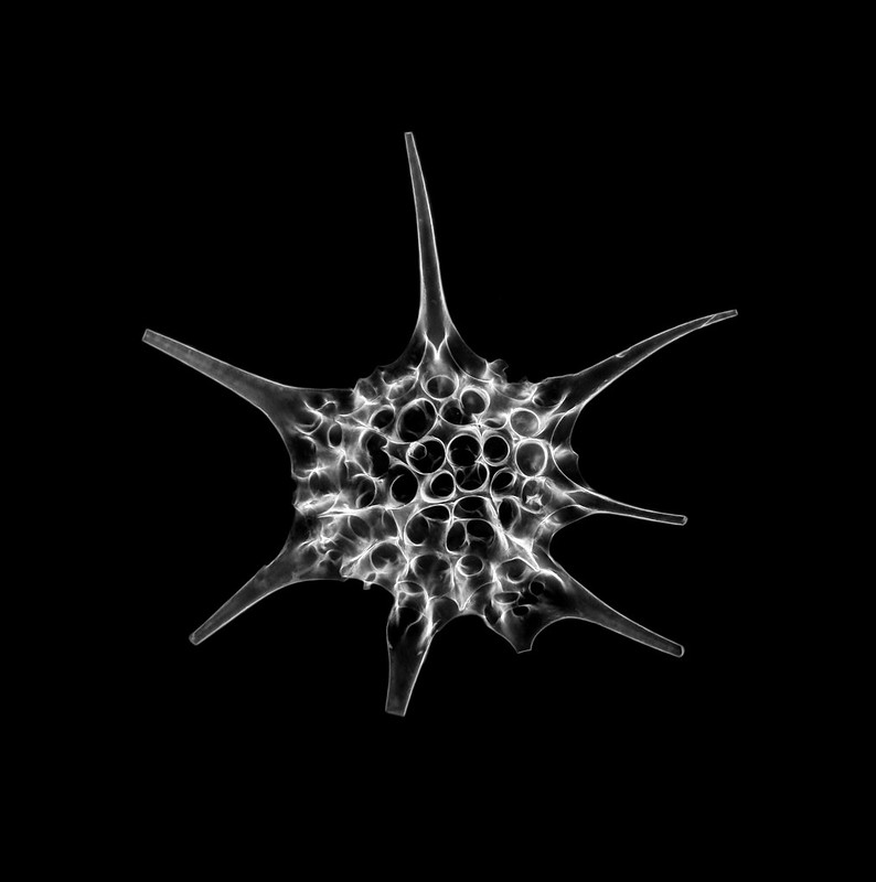
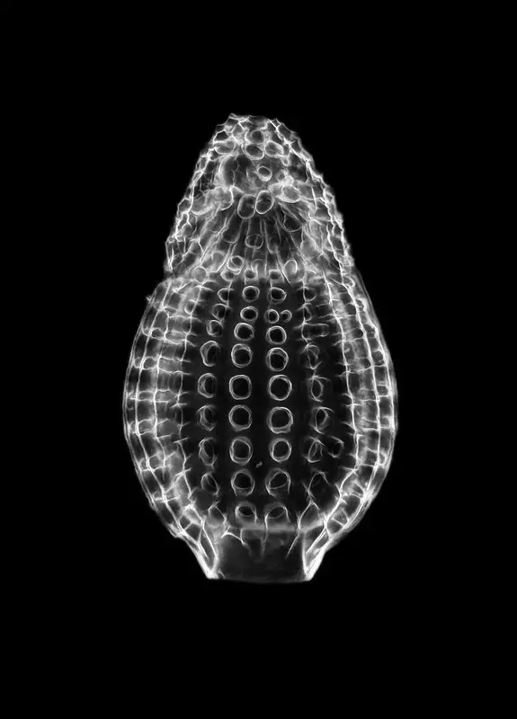
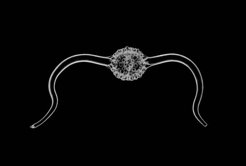
Radiolarians species, members of the subclass Radiolaria, are single-celled eukaryotes commonly found in marine environments (with some being colonial).
Although some of the species are restricted to a specific region, these organisms are widely spread in major oceanic ecosystems across the world.
Despite being closely related to amoebas, the majority of radiolarians are immotile and therefore incapable of movement.
Like Foraminifera, Radiolarians are characterized by shells that can be found in plenty of zones of high productivity (where they reproduce in high numbers). For the most part, Radiolarians are free-living organisms that feed on a variety of food sources in their environment. In the event of food scarcity, however, some of the species have been shown to benefit from symbiotic relationships with other organisms in their surroundings.
* Colonial Radiolarians include members of the families, Collozoida, Sphaerozoida, and Collosphaeridae.
Some of the species that fall under the subclass Radiolaria include:
- Acanthosphaera actinota
- Acrobotrys cribosa
- Acrosphaera cyrtodon
- Acrosphaera australis
- Acanthodesmia vinculata
- Acrosphaera spinosa
Classification of Radiolarians (based on the system proposed by Riedel, 1967)
Kingdom: Protista - The kingdom Protista consists of simple, eukaryotic organisms that are not plants, animals, or fungi. They exist in various environments across the world (aquatic, terrestrial, etc) and may exist as single cells or in colonies
Phylum: Sarcomastigophora - The phylum Sarcomastigophora consists of a variety of unicellular (occurring as single cells) and colonial organisms that move from one point to another with the use of flagella or pseudopods (or both in some organisms). While the majority of these organisms are free-living, some are parasites of both plants and animals
Subphylum: Sarcodina - Sarcodina is one of the largest phyla consisting of amebas and other organisms related to amoebas. In their habitats, these organisms may exist as single cells or in colonies with some of the species living as parasites. They are characterized by temporary cell extensions (pseudopods) that are used for movement and feeding.
Class: Actinopoda - Members of this class are characterized by tentacles that originate from the radial ambulacral vessels. Unlike many members of the subphylum Sarcodina, they are freely floating and therefore depend on water currents etc to move from one area to another.
Subclass Radiolaria
Ecology of Radiolarians Species
As mentioned earlier, Radiolarians species are widely distributed in major oceans across the world. In this environment, Radiolarians can be found at various depths, from the ocean surface to the bathypelagic depths (1000 to 3000 meters below the ocean surface).
Although the majority of species are found in marine environments, studies have discovered that Lophophaena rioplatensis thrive in brackish waters. In this environment, however, they only survive under certain conditions (salinity level of about 15.4 PSU). For this reason, they are absent in some areas of this environment (e.g. where salinity is below ca. 30 PSU).
* Brackish water is the type of water that has more salinity than freshwater but less salinity compared to seawater.
As a result of varying environmental conditions (salinity level, temperature, etc), some species are restricted to specific regions. Some Radiolarians serve as indicators of water properties and thus provide information regarding these environments.
Symbiotic relationships between Radiolarians species and algae are also dependent on their geographic location. For instance, Radiolarians that dwell in great depths live in areas that lack or have very little sunlight. For this reason, they lack algal symbionts given that algae cannot produce under such conditions (lacking light).
* The distribution of Radiolarians in various habitats can be determined by studying the number of Radiolarian skeletons deposited in different locations.
Characteristics of Radiolarians
Cell Ultrastructure
Spumellaria and Nassellaria are some of the most common Radiolarians species. They are characterized by a spherical body that consists of a centrally located nucleus (large in size with a complex shape). As microzooplankton, the majority of Radiolarians are very small in size, ranging from 30 to 300um.
The inner cytoplasm is highly organized compared to that of other related organisms and is separated from the outer part of the cell by an organic wall. The outer cell layer, which is very complex, consists of various cell organelles, inclusions (bubble-like inclusions), as well as a network of radiating pseudopods. For all species, some of the pseudopods differentiate to form axopods which consist of microtubules and a surrounding cytoplasm envelope.
Apart from the axopods, Radiolarians are also characterized by shells. In some of the species, these shells may be partially embedded in the cell substance and therefore does not completely cover the cell.
For the majority of Radiolarians, the shell extends the cell body and is normally covered by a thin layer of protoplasm. During the formation of this skeleton, the organism secreted amorphous opaline silica into the intracellular vesicles over time thereby gradually building the shell.
* Depending on the species, the skeleton ranges from 40 to 400um in size.
* Outside the cytoplasm, amorphous silica is deposited within an enclosing cytoplasmic sheath known as the cytokalymma. Cytokalymma is a dynamic sheath that plays an important role in molding the shape of the deposited silica during silification as the structure continues growing. According to researchers, this process is under genetic control and species-specific.
Compared to other organisms, such as Foraminifera species, the shells (exoskeleton) of Radiolarians are characterized by numerous spines that extend outward. However, they are also highly diverse and are often used as the basis on which different groups are grouped.
The outmost skeleton, also known as the cortical shell, is formed from the fusion of the spines. This shell is then connected to the inner shells by elongated spines/beams that provide structural support to the entire structure. The shell is also characterized by perforated holes (pores) through which pseudopods extend during feeding.
Among the Radiolarians species, feeding is also supported by axopodia. Unlike the pseudopodia, axopodia (long and slender) are supported by rigid central rods consisting of microtubule shafts. Being a more rigid structure, axopodia plays an important role in supporting the pseudopodia during feeding.
Food Sources
In their environment, Radiolarians feed on a variety of food materials. Using pseudopods and axopods, they trap and feed on such organisms as bacteria, diatoms, and small protists among other organisms. While some of the species are omnivorous and therefore eat both plants and tiny organisms in their surroundings, others are more specialized and only feed on certain prey.
In addition to simply depending on the food sources in their environment, some of the Radiolarians species have been shown to form a symbiotic relationship with algal. This is particularly common among Radiolarians that live on the upper layers of the ocean where sunlight is available for photosynthesis.
Here, the algal symbionts come in contact with the organisms and reside in the peripheral cytoplasm surrounding the central body of the cell. Given that they are exposed to sunlight, the algal symbionts manufacture their own food using water and carbon dioxide that supports the Radiolarian host when other food sources are not available.
Radiolarians are also an important food source for a number of organisms in their environment. They provide nutrition for such organisms as salps. As such, they are part of the food chain in their respective habitats.
* While prey may be trapped by pseudopodia, they may also be trapped on to the sticky surface of the axopodia before being engulfed through the invagination of the membrane. This results in the formation of an intracytoplasmic vacuole that is in turn converted to a digestive vacuole following the secretion of digestive enzymes.
Reproduction and Life Cycle
While some research studies have observed the production of gametes by Radiolarian cells, reproduction among these organisms is not properly understood. Among polycystines, a group of Radiolaria in the old classification, reproduction starts with the contraction of the extracapsular cytoplasm. At the same time, symbionts and waste matter are ejected.
Within the cell, the nucleus goes through several cycles of division which results in multiple nuclei filling the intracapsular cytoplasm. The nuclei, which are known as swarmers at this stage escape the cell through ruptures on the capsule wall. Given that fusion between swarmers has not been observed, their fate is not well understood.
Some of the main components of swarmers include:
- An organic envelope
- Vacuolar-bound strontium sulfate
* According to some studies, swarmers are first formed from the central capsule which then sinks to lower water depths causing it to rapture and release flagellated cells. Recombination of these cells then results in diploid adults.
* Most species are suspected to live for a few weeks or month .
Size and Colonies
Some Radiolarians species are large in size and can grow to between 1 and 2mm in diameter. These species are fewer compared to the other species and are often found on the water surface. Noncolonial species may lack the outer skeleton, but are enclosed by a gelatinous coat. However, some of them secrete siliceous spicules in the peripheral cytoplasm.
While many of the species exist as single cells, some form colonies consisting of numerous cells. Normally, these cells are interconnected by a network of cytoplasmic strands in addition to being enclosed in a gelatinous envelope that is produced by the cell.
Depending on the species, these colonies vary in size ranging from a few centimeters to over a meter in length. The shape of these colonies has also been shown to vary from spherical to ellipsoidal with some of the species forming elongated ribbon-shaped colonies.
Radiolaria vs. Phaeodaria
Traditionally, the subclass Radiolaria was divided into two main groups including Polycystines and phaeodarians. However, based on genetic studies, Phaeodarea (now classified under the class Thecofilosea) was shown to be a close relative of Radiolaria but not a group of the subclass. Being closely related, they share a number of characteristics but also vary in a number of ways.
Like members of the subclass Radiolaria, phaeodarians also have elaborate skeletons consisting of silica. Unlike Radiolarians, phaeodarians do not have axopodia. However, they produce a network of finely, interconnected pseudopodia that originate from the protoplasmic strands in the central capsule.
They are also larger in size compared to Radiolarians but do not normally form colonies. While they are also found in marine environments, phaeodarians are often found in deeper levels (below 300 meters).
Some of the other characteristics associated with phaeodarians include:
- Capsular wall that surrounds the endoplasm lacks fusules
- Skeleton may consist of materials other than silica - Various organic matter
- In some species, the skeletal framework consists of hollow tubes
- Sexual reproduction has not yet been observed
- Are closely related to testate amoebae
Sample Collection
Collecting living Radiolarians species is a simple process that may involve using a collecting jar or tow nets. While tow nets can be bought online, it's also possible to make one at home with the right material (a metal ring, metal rings, a nylon stocking, rope, and a bottle). Using the tow net or a jar, the specimen can be collected in areas where a high number of Radiolarian shells are found or where the organisms are known to be present in high concentrations.
Radiolarians Microscopy
* The specimen can first be sieved from the water before being placed on a microscope slide for viewing.
Requirements
- Compound microscope
- A sieve (45um sieve)
- Microscope glass slides
Procedure
· Using a sieve, sieve the water in order to retain the specimen (radiolarian)
· Try to obtain the sample and place it on a microscope slide - Some species are large enough to view with the naked eye (about 2mm) and can , therefore, be easily isolated. On the other hand, you can sieve the water and add small drops of water on a glass slide using a dropper for smaller organisms. This may take more time, but it is effective after sieving
· Observe the slide at ×100 and then using ×400 and record your observation
Isolating Radiolarians from Indurated Sediments
To obtain Radiolarian skeleton from quaternary sediments for observation, the following procedure can be used:
- Boil the sample in water - To disintegrate it
- Sieve using a 45um screen - This is aimed at removing tiny organic matter and other fine material
- Boil the residue and then add hydrogen peroxide to the water in order to prevent the sample from boiling over
- Sieve for a second time using the 45um screen
- To dissolve the carbonate, add diluted HCL (hydrochloric acid)
Observation
To observe the sample under the microscope, place the residue on a clean glass slide and allow it to dry. Embed the sample using Canada Balsam and cover it with a coverslip. Lastly, place the slide on the microscope stage and observe under ×100 and ×400 magnifications to record your observation.
Under ×400 it's not possible to view the cell body. However, the outer skeleton as well as different patterns and spines will be clearly visible given that they cover/surround the cell body.
Return from Radiolarians Species to MicroscopeMaster home
References
Arthur S. Campbell. (1952). An Introduction to the Study of the Radiolaria.
David Lazarus. (2005). A brief review of radiolarian research.
Demetrio Boltovskoy, Roger Anderson, Nancy Correa. (2017). Radiolaria and Phaeodaria.
Kjell R. Bjørklund. (2016). Radiolarians.
Links
https://www.radiolaria.org/quaternary.htm
https://www.ucl.ac.uk/GeolSci/micropal/radiolaria.html
Find out how to advertise on MicroscopeMaster!


