Structure and Functions of
Cilia and Flagella
Overview
Cilia and flagella are fine, whiplike/hairlike structures that extend from the body of a variety of cells. While they vary in terms of length and numbers in different types of cells (as well as patterns of movement), cilia and flagella are generally identical in structure and composition.
Depending on the type of cells, cilia and flagella have the following functions:
· Propelling cells - Using cilia or flagella, cells are able to move freely in their environment, especially in aquatic or moist environments.
· Sensory functions - Some cilia and flagella allow cells to sense changes in their surroundings which in turn allows the cells to respond appropriately.
· Transporting material - Some cells are able to not only trap, but also guide the transportation of given material. This may serve to engulf such material into the cell or prevent unwanted material/particles/microorganisms from invading the cell or tissue.
* The flagella of prokaryotes have a different structure compared to those of eukaryotic cells.
Cilia
With the exception of a majority of higher plants and fungi, cilia can be found on the surface of many eukaryotic cells. On these cells, cilia extend from the basal body.
Depending on the type of cells, cilia have several functions and are therefore divided into two main categories.
* Prokaryotes (bacteria) do not have cilia.
Cilia Structure
Cilia are microscopic, hair-like structures that project from the surface of many eukaryotic cells. Like other organelles of eukaryotic cells, cilia are membrane-bound structures with their membrane being continuous with the plasma membrane.
Unlike the plasma membrane of cells, ciliary membrane has been shown to contain distinct lipids and proteins.
Motile Cilia
* Motile cilia were identified in the 1640s by van Leeuwenhoek making them the earliest known cell organelles.
Motile cilia (9+2) can be found in both higher animals and single-celled eukaryotes. In microscopic organisms (known as ciliates) motile cilia are used for locomotion or for moving fluid over their surface which contributes to the feeding process.
In higher animals, such as human beings, motile cilia can be found in a number of tissues (e.g. respiratory epithelium and fallopian tubes) where they are either involved in the clearance of or moving of substances.
In the respiratory system, cilia trap and remove dirt (as well as mucous) from the lungs and other parts of this system. In the fallopian tube, on the other hand, cilia serve to move the ovum to the uterus.
On the cell surface, motile cilia are present in large numbers where they beat in a coordinated wavelike manner to perform their functions effectively.
Some examples of ciliates include:
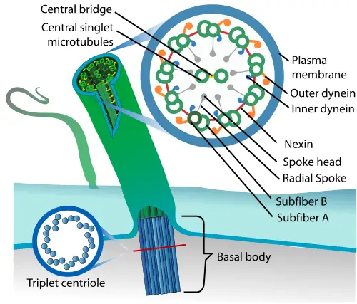
With regards to structure, motile cilia are characterized by a radial pattern consisting of nine (9) outer microtubule doublets that surround two singlet microtubules.
Here, then, the 9+2 pattern refers to the nine doublet microtubules surrounding the two microtubules that are centrally located. The ring of microtubule scaffolding, in this case, is known as the axoneme.
In addition to the microtubules, which are the main components of the structure, motile cilia are also composed of dynein arms and radial spokes that contribute to the overall motility of the structure.
* The axoneme (the bundle of microtubules which measures about 0.25um in diameter) is surrounded by the plasma membrane and the whole structure (cilia) can be identified under the microscope.
At its base (where it attaches to the cell), the axoneme is attached to cylindrical structures known as basal bodies. The basal bodies measure about 0.4um in length and 0.2um in width and are made up of the A tubule (nine (9) triplet microtubules consisting of protofilament microtubules), an incomplete B tubule as well as an incomplete C tubule.
Apart from anchoring cilia in the cytoplasm, basal bodies also play an important role in the assembly of these structures.
* According to studies, basal bodies are either the products of centrioles or are generated in large numbers before cilia formation.
* Even when the surrounding plasma membrane has been removed, the addition of ATP allows the axoneme continues to function which is evidence that the working mechanism of the structure resides in the axoneme.
Beating Mechanism of Cilia
As is the case with muscle contraction, the beating/working mechanism of cilia (axoneme in particular) has been shown to be the result of sliding protein filaments.
Although the mechanism, in its entirety, is yet to be fully understood, studies have shown dyneins, which act as the molecular motors, to play an important role in powering the ciliary beat.
One of the models that have been used to describe the bending and thus the functioning of motile cilia is the switch model hypothesis.
According to the switching model, each side of a curved cilia consists of dyneins in a given state of force generation cycle which contributes to the asymmetry and change with alterations in curvature.
Here, according to studies, dyneins on one microtubule (in the force generation cycle state) slide past each other while those on the other side do not. This results in the bending of the axoneme while the switching of this system causes the structure to bend to the other side.
Ultimately, the repeat of this mechanism causes motile cilia to beat and thus perform their function.
* The attachment and release of dynein arms to adjacent doublet is caused by binding or hydrolysis of ATP.
Primary Cilia (Non-Motile Cilia)
As compared to motile cilia, primary cilia (9+0) project as single structures from cell bodies. They are found in virtually all cells in all mammals. They are primarily involved in sensory functions and thus allow given body tissues/organs to respond appropriately.
Like motile cilia, primary cilia consist of nine doublet microtubules that make up the axoneme. These microtubules originate from the basal body that also provides stability.
Unlike motile cilia, however, primary cilia do not possess dynein arms and the central singlet microtubules (central pair microtubules). This is due to the fact that primary cilia are not motile and thus do not need elements necessary for motility.
* Initially, primary cilia were thought to be vestigial organelles that served no purpose.
Formation of Primary Cilia
Primary cilia formation begins when a cell enters the G0 phase of the cell cycle. Here, the mother centriole of the centrosome first attaches to the vesicle followed by the growth of the axoneme from the surface of the centriole.
Axonemal microtubules also start to polymerize at the growing tip where cargo is delivered through intraflagellar transport.
This bidirectional transport system allows for proteins to be transported to the microtubules during its development. While the vesicle is ultimately exocytosed, the primary cilia are exposed at the surface of the cell and continue to develop until it reaches maturity.
Intraflagellar transport (IFT) is still necessary for the maintenance of primary cilia.
* Primary cilia have been shown to align in a single direction which in turn influences the orientation of cells.
Functions of Primary Cilia
Primary cilia play an important role in cell signaling during development and homeostasis. Given that primary cilia (5-10um in length) are exposed to the extracellular environment, they are susceptible to various stimuli that contribute to their role in signaling.
In addition to detecting various chemical factors, morphogens and growth factors in the extracellular matrix, primary cilia also detect changes in pressure and fluid movement across the cell surface.
For instance, due to the flow of urine in the kidney tubules, primary cilia are influenced to bend which in turn results in the influx of calcium ions through appropriate calcium channels.
Apart from Hedgehog pathways, Wnt signaling pathways is one of the best-documented pathways with regards to ciliary signaling. Essentially, Wnt signaling pathway is important because it is involved in a number of processes including cell polarity, cell migration as well as neural patterning among others.
It occurs in two major paths including the canonical Wnt pathway and the non-canonical Wnt pathway.
Whereas the activation of the Canonical Wnt pathway contributes to gene expression, the activation of the non-canonical Wnt pathway results in the degradation of b-catenin. Here, the binding of various Wnt ligands to receptors located on primary cilia allows canonical Wnt signaling to switch to non-canonical signaling and vice-versa.
The role of primary cilia is also evident in a number of other signaling pathways allowing for appropriate responses. Defects in cilia functions, on the other hand, have been associated with various developmental and degenerative diseases.
Defective cilia functions have been associated with the following disease and disease syndromes:
- Primary cilia dyskinesia
- Alstrom syndrome
- Meckel-Gruber syndrome
- Nephronophthisis
- Respiratory infections
- Anosmia
- Male infertility
Flagella
A flagellum (plural: Flagella) may be described as a filamentous organelle that is primarily used for locomotion. Like cilia (found in eukaryotic cells), flagella also protrude from the body of the cell which allows them to perform their functions effectively. However, they are longer in length, measuring between 5 and 20um.
Cells that possess this structure are referred to as flagellates and include both eukaryotic and prokaryotic cells. For instance, apart from a majority of bacteria that use flagella for locomotion, the structure can also be found on such single-celled organisms as euglena and protozoa species like Trypanosoma evansi. Flagella, also, lagella can be found on gametes of various organisms including slime molds, fungi, and animals.
Flagellum Structure
While flagella can be found in both eukaryotic and prokaryotic cells (and serve the same purpose) there are various differences with regards to their structures/composition as well as the mechanism by which they function between the two types of cells.
The flagella found in prokaryotic cells consist of a globular protein known as flagellin. Here, the protein wraps around in a helical manner forming a hollow cylinder along the length of the structure. This protein is absent in eukaryotic flagellum where it's replaced by protein filaments known as microtubules.
Some of the other differences between the two include:
- Prokaryotic flagella tend to be smaller and less complex compared to eukaryotic flagella
- Eukaryotic flagella are powered by ATP while those of prokaryotes are proton-driven
- The prokaryotic flagella are characterized by a rotator movement while those of eukaryotic cells have bending fashion
- Prokaryotic flagella lack a plasma membrane
Apart from length, the structure and composition of eukaryotic flagella are similar to cilia found in many eukaryotes (described above).
This section will, therefore, focus on the structure of flagella found in prokaryotic cells.
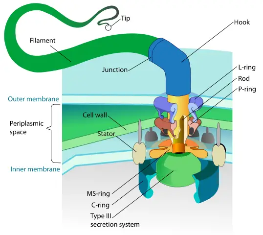
Bacterial flagellum is composed of three main parts that include:
- Basal structure (Rotary motor)
- Hook (acts as the universal joint)
- Filament (the helical propeller)
Basal body - In bacteria/prokaryotes, the basal body is a rod that consists of several rings that are located within the cell membrane. In Gram-negative bacteria, the rings include the L-ring that is positioned in the outer membrane of the lipid bilayer and the P ring which is located in the peptidoglycan layer.
The basal body is generally divided into several parts that include:
- Protein rings (C ring, MS ring, P ring, and L ring)
- Rod
- Sleeve
* Protein rings serve as the proton pumps that are involved in the movement of hydrogen ions across the membrane. It's this movement of ions across the membrane that ultimately rotates the rings and thus the flagellum.
* The basal body, as well as the hook, also serves to anchor the filament of the structure to the surface of the cell.
The Hook
Consisting of 120 subunits of a single protein, the hook (which is short and curved) acts as the universal joint that connects the filament to the basal body.
Unlike the basal body, the hook is not embedded in the plasma membrane. However, it plays a crucial role in the motility and taxis of bacteria through the transmission of motor torque to the filament (propeller) part of the structure.
It's composed of 4 main domains that are arranged on the inside and outside of the structure whose nature allows for the direct connection between the hook and the rod.
* The junction between the hook and the filament consists of two proteins (FlgK and FlgL) which have been shown to contribute to the formation of the filament part of the structure.
The Filament
The filament is the elongated part of the flagella. It's tubular and consists of 11 protofilaments that resemble those found in the rod and hook parts of the structure.
The flagellin, which is the main component of the filament, also consists of four domains that form the inner and outer part of the structure. The direction to which filament rotates is dependent on the motor spinning (clockwise or counterclockwise).
While flagella found in such organisms as bacteria, archaea, and various eukaryotic cells are used for swimming through fluid as well as swarming; they have also been shown to serve a number of other important functions. For instance, in eukaryotic cells, the structure has been shown to play a role in increased production.
In both bacteria and eukaryotic cells, some flagella have been shown to have sensory functions that allow cells to detect changes in their environment and respond effectively. In some green algae, studies have suggested that flagella may act as secretory organelles.
* Organisms may be classified based on the number of flagella on their surface.
These include:
- Monotrichous - single flagellum originating from one end
- Lophotrichous- several flagella at one pole
- Amphitrichous - single flagellum on both poles
- Peritrichous - multiple flagella across the surface of their bodies
Return from Cilia and Flagellum to MicroscopeMaster home
References
Brent W. Bisgrove and H. Joseph Yost. (2006). The roles of cilia in developmental disorders and disease. The company of Biologists.
Hiroyuki Terashima, Seiji Kojima, and Michio Homma. (2008). Flagellar Motility in Bacteria:
Structure and Function of Flagellar Motor. International Review of Cell and Molecular Biology, Volume 270.
Takashi Ishikawa. (2017). Axoneme Structure from Motile Cilia.
Stephen M. King. (2018). Turning dyneins off bends cilia.
Witman G.B. (1990) Introduction to Cilia and Flagella. In: Bloodgood R.A. (eds) Ciliary and Flagellar Membranes. Springer, Boston, MA.
Yuko Komiya and Raymond Habas. (2008). Wnt signal transduction pathways. ncbi.
Links
https://www.researchgate.net/publication/235374058_Cilia_Wnt_signaling_and_the_cytoskeleton
https://academic.oup.com/bioscience/article/64/12/1073/250556
Find out how to advertise on MicroscopeMaster!
![Beating pattern of flagellum and cilia, Original: Kohidai, L.Vector: Urutseg [CC BY 3.0 (https://creativecommons.org/licenses/by/3.0)] Beating pattern of flagellum and cilia, Original: Kohidai, L.Vector: Urutseg [CC BY 3.0 (https://creativecommons.org/licenses/by/3.0)]](https://www.microscopemaster.com/images/512px-Flagellum-beating.svg.png)


