Phylum Sarcodina
** Examples, Classification and Characteristics
Overview
Classified as a subphylum or superclass in some literature, phylum Sarcodina consists of amoeboid protozoa that move and feed by means of cytoplasmic streaming. The majority of species can be found in aquatic environments where they exist as free-living organisms. However, a few species are parasitic and cause harm.
Phylum Sarcodina is divided into two major groups that include:
Classification
Kingdom: Protista
Subkingdom: Protozoa
Phylum: Sarcodina
Superclass: Rhizopoda - Classes Lobosa, Acarpomyxea, Eumycetozoea, Granuloreticulosea, Filosea, and Plasmodiophorea
Superclass: Actinopoda - Classes Radiolaria, Heliozoea, and Phaeodarea.
* Phylum Sarcodina is estimated to consist of over 12,000 species.
Phylum Sarcodina: Characteristics of Superclass Rhizopoda
Lobosa (Lobosea)
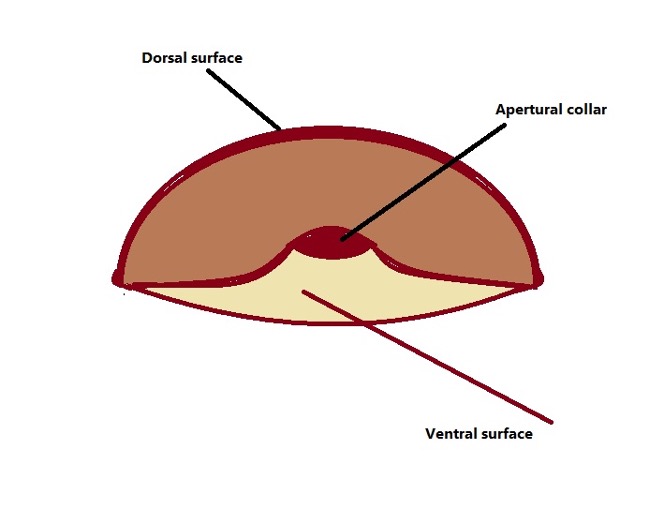
Morphology - The class Lobosa consists of testate and naked amoeba. Arcella and Difflugia species are examples of shelled amoeba in this group. Arcella species are characterized by an umbrella-shaped shell of varying sizes depending on the species. These shells are organic in nature and may contain pores.
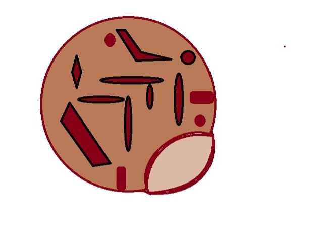
As the amoeba grows, the shells accumulate iron and manganese which causes them to become brownish in color. Difflugia species, on the other hand, are characterized by more rounded/ovoid shells/tests. These shells consist of a variety of minerals collectively known as xenosomes. Naked species lack shells and therefore do not have a constant general body form. Here, some of the species include A. proteus, A. amazonas, and A. borokensis.
Depending on the species, the cells are binucleate or multinucleated. They can form filiforms, lobopodia, and subpseudopodia for motility. For instance, Arcella species produce lobopodia which emerges from the aperture of the cell.
* Difflugia species vary in shape, size, and color
* Some species (e.g. some members of the genus Acanthamoeba) form cysts during unfavorable conditions.
Ecology/Habitat - Lobosa species are commonly found in wet habitats including moist soils, plants in aquatic environments, freshwater sediments, and wet foliage etc. These environments are essential in that they prevent the cells from drying out and also act as sources for various minerals required to build the shells.
Nutrition - In their respective habitats, Lobsa species feed on fungi, algae, and bacteria. This is achieved through a process known as phagocytosis. Here, the amoeba extends the pseudopods (cytoplasmic extensions) which allow them to engulf food material and digest them within the cell.
Examples of Lobosa species:
- Spongia lobosa
- Naegleria species (E.g. N. gruberi, N. Jowlen)
- Acanthamoeba species (e.g. A. castellani, A. palestinensis)
Acarpomyxea
Morphology - Members of the class Acarpomyxea are generally small in size. Some have been shown to exist as unicellular plasmodia while the majority is branched amoebae. The pseudopods move and feed by means of the cytoplasmic extensions (pseudopodia) which also consists of microfilaments.
Habitat - The majority of species can be found in moist soil, coral reefs, and some aquatic environments. Here, they have been shown to feed on diatom protoplasts.
* A few species are suggested to be multinucleated.
* They contain granular cytoplasm.
Examples of Acarpomyxea species:
- Paraflabellula reniformis
- Leptomyxa reticulata
- Leptomyxa reticulata species
Eumycetozoea
Morphology - Eumycetozoea species are unique from some of the other Sarcodina in that they form filiform subpseudopodia. Compared to other pseudopodia, filiform subpseudopodia are shaped like filament and are thus described as thread-like.
Some of the species have also been shown to produce flagella for locomotion (swimming). In some of the species, fruiting bodies with elongated stalks tubes are also produced. Also known as slime animals, Eumycetozoea cells contain semifluid protoplasm in which nuclei are situated.
The flagellated forms (plasmodia) are produced by spores during favorable environmental conditions and undergo sporulation when conditions deteriorate.
Habitat - They can be found on some aquatic plants where they survive by feeding on bacteria among other tiny organisms.
Examples of Eumycetozoea species:
- Licea species
- Trichium species
Plasmodiophorea
Plasmodiophorea consists of genera like Spongospora, Woronina, Ligniera, and Plasmodophora, etc. While their species are classified under the group Rhizopoda, they are known considered true protozoa.
Morphology and life cycle - Members of this group are collectively known as plasmodiophorids and exhibit significant variation in morphology when compared to other protozoa. With the exception of a few species, the majority of species in the group can form resting spores known as cystosori.
Under conducive conditions, these resting bodies give rise to amoeboid cells. They are also distinguished by several important characteristics that include a chitinous cyst wall, Golgi bodies in plasmodia, and the absence of phagotrophy.
As mentioned, their life cycle consists of several stages that include resting spores, zoospores, and amoeboid cells (sporangia). Here, the life cycle starts with the penetration of zoospores into plant root hairs. When they come in contact with root hair cells of the appropriate host, zoospores form a tubular structure known as Rohr that consists of a Stachel (elongated pointed body).
This is followed by the production of adhesorium for attachment and injection of Stachel and contents of the zoospore into the cytoplasm of the host cell.
Within the cell, these contents increase in size and undergo several stages of cruciform nuclear division giving rise to multinucleated plasmodia (sporangia). The sporangial plasmodium has a distinct cell membrane and cell wall that isolates them from the cytoplasm of the host cell.
Over time, a few septa are formed in the sporangium dividing it into lobes. This process gives rise to a zoosporangium which is larger in size.
The zoosporangium goes through several stages of mitotic nuclear division (non-cruciform) and forms tubes that extend through the cell wall of the host. These exit tubes allow secondary zoospores to be released into the environment (outside the plant root) or into the neighboring cells.
In the environment, they can transform to resting bodies or sporogenic plasmodia and sporangial plasmodium when they come in contact with the roots of host plants.
* In the root cells of the host plant, sporogenic plasmodia are characterized by a thin membrane that separates the organism from the cytoplasm of the plant cell. This differentiates them from sporangial plasmodia which have a cell wall separating them from the cytoplasm of the host cell. Moreover, sporogenic plasmodia are generally larger and thus fill the entire cross-section of the host plant cell.
Some of the other morphological characteristics of Plasmodiophorea include:
- Zoospores are uninucleate and biflagellate (flagella originate from the anterior part of the body)
- Flagella are used for motility
- They all form cysts (resting spores). These spores are characterized by a smooth or ornamented chitinous outer layer. In this form, they can survive in soil for many years
* In a few species, reproduction occurs through planogametic copulation. This is a form of sexual reproduction.
Habitat - Plasmodiophorea species are endoparasites of plant cells. As such, they are commonly found in the cells of plant roots. However, some of the species invade fungi and algae cells. In the absence of the host, they can be found in the soil as cysts (resting cells). As parasites, they can cause significant damage to the plants resulting in high losses.
* Some species are hosts of viruses.
Examples of Plasmodiophorea species:
- Polymyxa graminis
- P. betae
- P. brassicae
Filosea
Morphology, Life Cycle, and Habitat
The genus Nuclearia is one of the best-studied groups within the class Filosia.
Although they do not necessarily produce cysts, their life cycle consists of a cyst-like stage characterized by short pseudopodia that originate from one end of the cell body.
The trophic forms, on the other hand, tend to be spherical in shape and range between 22 and 50 um in diameter. These forms produce filosea pseudopodia which are short and thick. Aside from filose pseudopodia, they also produce contractile pseudopodium.
Depending on the species, the cells are multinucleated with an average of 12 nuclei. Some of the other organelles identified under the microscope include vacuoles, mitochondria with flattened cristae, and dictyosomes. Most of the species can be found in marine environments in association with various animals (e.g. fish). However, some of the species (e.g. some Chromatophora) can be found in freshwater or brackish water.
* Some of the species in this group (e.g. Paulinella species) have a shell for protection.
Granuloreticulosea
Morphology and Habitat
The majority of species in the class Granuloreticulosea are characterized by mineralized shells. These shells may have one or more apertures. Some of the species do not have a shell and produce branched rhizopodia. The cell body contains multiple nuclei, granular cytoplasm, food vacuoles, mitochondria, and Golgi bodies, etc. A few of the species produce amoeboid gametes some of which are biflagellate.
They are commonly found in marine habitats and brackish water where they feed on diatoms, algae, bacteria, and other small prey.
* The majority of species in this group are fossils.
Examples of Filosea species:
- N. moebiusi
- N.delicatula
Phylum Sarcodina: Characteristics of the Superclass Actinopoda
The majority of species in the Superclass Actinopoda in phylum Sarcodina are free-floating organisms found in aquatic habitats.
They include:
Radiolarians
The term Radiolaria is used to refer to large marine Actinopoda that float and trap their food (plankton) using rhizopodial networks.
Morphology
Like many other species in phylum Sarcodina, Radiolarians are single-celled protozoa. As mentioned, they are large in size, ranging from about 40um to 400um in diameter.
While the smaller ones can only be observed under the microscope, larger ones can be viewed with the naked eye (larger species are less numerous than smaller ones). They contain a glass-like exoskeleton made up of silica material obtained from the environment.
Here, the silica materials obtained from the environment (aquatic environment) are developed into geometric networks to build the skeleton/test. These tests are also characterized by spines and have pores that allow pseudopodia (rhizopodial networks) to extend.
While Radiolarians are skeleton-bearing organisms, the morphology of these skeletons varies from one species to another. Among the larger species, cells might be enclosed in a gelatinous covering. Compared to many other Radiolarians, these species do not have a skeleton. Others are enclosed in siliceous spicules scattered within the gelatinous layer.
Among the smaller species (capable of forming colonies), the cells are interconnected by strands of cytoplasm within a gelatinous coating. These colonies vary in size from as small as a few centimeters to over one meter in length.
Apart from variations in size, these colonies also vary in shape from spherical and ellipsoidal to cylindrical and ribbon-shaped. These differences have proved useful for identifying the different species within the group.
* A colony of collodaria may comprise hundreds to thousands of cells held together by a gelatinous matrix.
* Some species produce small flagella.
* The cells of radiolarians have a central capsule between the endoplasm and ectoplasm. This is unique to these organisms.
Habitat and Nutrition
The majority of Radiolarians are marine organisms widely distributed in Oceans across the world. Here, they are largely concentrated in warm areas around the equatorial zone from the ocean surface to several hundred meters deep.
For the species found on the surfaces, microscopic studies have revealed numerous air vacuoles that allow them to remain afloat. In world Oceans, they feed on a wide variety of prey and material including diatoms, organic detritus, ciliates, copepods, and certain flagellates, etc.
Reproduction - Generally, Radiolarians reproduce asexually through binary or budding. Here, the parent cell gives rise to a daughter cell during budding or two new daughter cells during binary fission. As well, sexual reproduction has been reported in some species.
Some members of the class Radiolaria include:
- Taxopodia
- Polycystinea
- Acantharia
Heliozoea
Morphology
Like Radiolarians, some Heliozoans are testate organisms. Their tests/shells are made up of silica among other organic materials (e.g. gelatinous substance).
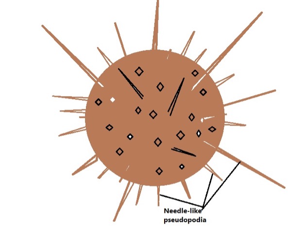
Though some of the species have perforated shells, they lack an organic lining between the central mass and cytoplasm found in some of the other members of the phylum Sarcodina. However, they may have a thin mucous coating over the cell membrane. They are characterized by a spherical body and produce cytoplasmic projections known as axopodia used for motility, capturing prey, and adhesion.
Some of the species have also been shown to produce fine pseudopodia. The cell body consists of a granular cytoplasm in which the nucleus, contractile vacuoles, and other organelles are located. In some of the species (e.g. Actinosphaerium eichhorni), the cell contains many nuclei that range between 5 and 10 um in diameter.
Habitat and nutrition - Unlike Radiolarians, Heliozoans are commonly found in freshwater habitats. Here, they use their cytoplasmic projections to capture prey which includes bacteria and crustacean larva, etc.
* The axopodia produce paralyzing substances that paralyze prey.
Some members of this group include:
- Clathrulina elegans
- Hedriocystis reticulata
- Actinosphaerium eichhorni
- Echinosphaerium nuclolfilum
Phaeodarea
* Members of the class Phaeodarea were previously classified under the class Radiolaria.
Morphology
Phaeodarea species are characterized by a skeleton composed of hollow silica spines. These spines are connected to each other by an organic material secreted by the organism. The cell body is of average size, ranging between 100 and 500 um in diameter. They produce axopodia which radiate from the cell body and through pores on the shell.
Like some of the other species of Sarcodina, they are uninucleate and thus possess a single large nucleus. The cell also consists of vacuoles and oil droplets among other organelles found in eukaryotic cells.
* Phaeodarea species have a unique central capsule as well as a vesicular body consisting of waste material known as a phaeodium.
* They also have a very thick capsular membrane that allows them to survive deep in the ocean.
Habitat and nutrition - Phaeodarea are commonly found in world Oceans below the photic zone where they feed on small plankton.
General Characteristics of Phylum Sarcodina
Generally, species in phylum Sarcodina are protozoa most of which feed through pseudopodial actions. While most of the species produce pseudopodia, these cytoplasmic projections vary in size and shape.
Whereas some of the pseudopodia are in the form of a single broad lobopodium, others are thin and numerous, radiating from all sides of the cell body. The shape of the cell body is largely dependent on the species. As mentioned, some of the species have a shell/test which influences the general shape. However, naked species often exhibit significant variation in shape.
Most Sarcodina species are free-living species found in various aquatic environments. In these habitats, they exist as carnivores and mostly feed by chance (they do not pursue prey).
Some of the most common prey include bacteria and crustacean larva, and other protozoa, etc. A few are herbivores and can survive on algae and other organic debris in their surrounding while some are parasitic and obtain their nutrition from the host (e.g. some Entamoeba species).
Depending on the species, feeding may occur through phagocytosis or pinocytosis (endocytosis). In both cases, food materials are engulfed and internalized into the cell where they are broken down by specific enzymes.
Waste materials are then released into the environment through exocytosis.
Phylum Sarcodina is often also classed as a subphyla under phylum Sarcomastigophora
What does phylum in biology mean?
Return from phylum Sarcodina to MicroscopeMaster home
References
Alexey V. Smirnov and Andrew V. Goodkov. (1999). An Illustrated list of basic morphotypes of Gymnamoebia (Rhizopoda, Lobosea).
Anderson O.R. (1988) Amoebae and Their Relatives (Phylum: Sarcomastigophora; Subphylum: Sarcodina). In: Comparative Protozoology.
Despommier D.D., Karapelou J.W. (1987) Protozoa. In: Parasite Life Cycles.
Nisbet B. (1984) Sarcodina. In: Nutrition and Feeding Strategies in Protozoa.
Roger Anderson. (1988). Comparative Protozoology: Ecology, Physiology, Life History.
Links
https://www.nies.go.jp/chiiki1/protoz/morpho/heliozoa/nucleari.htm
https://www.sciencedirect.com/topics/earth-and-planetary-sciences/radiolarian
Find out how to advertise on MicroscopeMaster!


