Phylum Rhizopoda
** Definition, Classification and Characteristics
***Also seen as a Superclass
Definition: What is Rhizopoda?
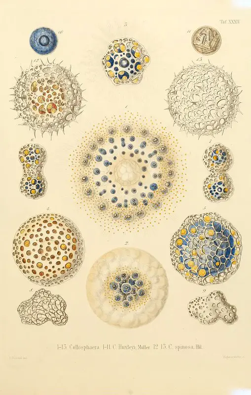
Although it was formerly ranked as a sub-class, Rhizopoda is today regarded as a phylum of the kingdom Protista. It consists of both naked and testate amoebae as well as some slime moulds and Foraminifera.
Like many other members of the Kingdom Protista, organisms classified under the phylum Rhizopoda can be found in various aquatic and terrestrial habitats across the world. They are characterized by possession of pseudopodia used for feeding and locomotion.
While the majority of species are free-living in their respective habitats, some are parasitic in nature and cause disease in human beings.
Some of the species belonging to the phylum include:
- Difflugia tuberspinifera
- C. puteus
- Cyclopyxis eurystoma
- Amoeba proteus
- Pseudothecamoeba proteoides
* While Rhizopoda is ranked as a class in some books, it's also ranked as a superclass in others.
* The name "Rhizopoda" means root feet.
Classification of Rhizopoda
According to the older method of classification, Rhizopoda was classified as follows:
· Kingdom: Protozoa - Single-celled eukaryotes that may exist as free-living organisms or as parasites
· Subphylum: Sarcodina - A large group consisting of amoebas and amoeba-like organisms that move and trap food with the use of pseudopods. This group also includes both free-living and parasitic organisms
· Superclass: Rhizopoda - A group consisting of naked and testate amoeboid organisms many of which use pseudopods to move and capture food material
· Classes: Filosia, Granuloreticulosea, Lobosa, and Xenophyophorida
* Rhizopoda is generally regarded as a separate phylum of the Kingdom Protozoa.
Characteristics of Rhizopoda
Diversity
While the phylum has also been shown to consist of several slime moulds and Foraminifera, it's mostly composed of naked and testate amoebae. Unlike naked amoebae, testate species are covered by a shell-like structure which may consist of small grains of sand, diatoms, mineral particles, and silica plates.
To make this structure, the single-celled organism identifies and collects the necessary material and then cements them around its body using its secretions. Good examples of these organisms include Arcellavulgaris, Centropyxis aculeata, and Centropyxis.
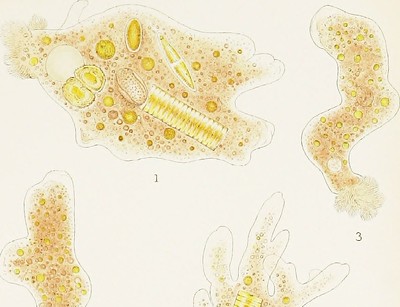
Naked amoebae, on the other hand, are not encased and are therefore described as being "naked". They mostly appear transparent and can be found attached to various surfaces in their environment.
Although they lack a test, naked amoebae have been shown to contain several rigid morphological features that allow for the identification of different species. Some of the species that represent this group include Amoeba flowersi and members of the genus Polychaos.
Members of the phylum Rhizopoda have also been shown to be very diverse in various habitats. Based on studies carried out in the 1990s, 46 testate species were discovered in terrestrial mosses in Hungary, 25 in forest moss in southeastern Alps in Italy and many others in aquatic environments across the globe.
While Rhizopoda species can be found in various environments across the world (moss, ponds, swamps, the Antarctic etc) they all require moisture to survive.
In cases where there is a lack of moisture (drought etc) many species have been shown to encyst in order to survive. Some of the species capable for forming cysts are Chlamydomyxa montana and Plagiomnium cuspidatum.
Testate amoebae are divided is divided into two groups that include:
Those with lobose pseudopodia
Lobose amoebae (testate) consist of various species that have been shown to be over 100um in size. As the name suggests, they are characterized by lobose pseudopodia known as lobopodia.
When viewed under the microscope, these projections appear to have a smooth outline and rounded tips. They are made up of cytoskeleton that consists of actomyosin filaments and plays an important role in movement (referred to as monopodial locomotion) and feeding.
Apart from these forms of pseudopodia, lobose amoebae have also been shown to produce structures known as subpseudopodia. Unlike the pseudopods with rounded tips, subpseudopodia are mostly hyaline projections with varying shape and do not result in the relocation of the cell mass.
Testate filose amoebae
Unlike testate lobose amoebae, testate filose amoebae are closely related to Foraminifera and are smaller in size (below 100um). Members of this group are also characterized by siliceous tests that contain idiosomes synthesized by the organisms.
Pseudopodia in these species are mostly composed of ectoplasm and have pointed ends.
Foraminifera
Like naked and testate amoebae, Foraminifera are also amoeboid protists. Members of this group are commonly found in deep-sea environments where they are some of the most abundant organisms.
They are characterized by a net-like system of pseudopodia that plays an important role in the feeding process.
Apart from the unique pseudopodia, they are also encased in a test/shell that may consist of one or several chambers. As is the case with testate amoebae, these shell-like structures are made up of particles like mineral grains among others held together by substances secreted by the organisms.
Some of the most common foraminiferans include Elphidium and Globigerina
* Apart from food gathering, the net-like pseudopodia, which are supported by sophisticated cytoskeleton (made up of microtubules), are also used for respiration, locomotion and construction of the shell-like test.
* Members of this group can grow to be several millimeters in length.
Nutrition/Feeding
A majority of Rhizopoda species are free-living organisms found in various aquatic and terrestrial environments. In these environments, Rhizopoda has been shown to heterotrophic with various species feeding on available organic matter.
Some of the species have been shown to form a symbiotic relationship with photosynthetic algae which allows them to obtain nutrition as they house these organisms while others feed on bacteria. Some of the other food sources of Rhizopods include plant cells, fungi, and smaller amoebae.
For free-living members of the phylum (both naked and testate amoebae), studies have shown them to move randomly in search of food. As compared to the naked forms, testate amoebae tend to change direction more abruptly.
Because of the manner in which they move about using pseudopodia (by crawling on surfaces) in search of food, this behavior of free-living Rhizopods is often described as grazing in various books.
When they come in contact with prey, they use pseudopodia to envelop it and enclose it in the food vacuole where it is digested. This process is particularly easy for naked, free-living Rhizopods given that they are not inhibited. In testate species, the shell-like covering gets in the way between the cell body and prey.
For this reason, some of the species are forced to rely on diffusion feeding. Here, projections protrude through pores/holes in the shell allowing the organism to capture prey. Here, the sticky nature of the cytoplasm has been shown to play an important role in capturing prey during feeding.
In some species, however, the pseudopodia protrude through a single opening and capture prey.
Parasitic Rhizopods
A majority of Rhizopods are free-living organisms commonly found in aquatic and moist terrestrial habitats. However, some members of this group are parasitic in nature and capable of causing diseases. As such, they can be found in the bodies of their respective hosts.
In human beings, Entamoeba histolytica is responsible for amebiasis. On the other hand, E. dispar, which is dependent on human hosts are harmless and do not cause diseases (they exist as harmless commensals).
The life cycle of E. histolytica starts when the cysts are ingested along with contaminated foods or water. In the small intestine, trophozoites are released from the cysts (as the touch wall is degraded by digestive enzymes) and migrate to the large intestine (colon) where they divide and multiply through binary fission (asexual reproduction). In the lumen, they have been shown to feed on bacteria and tissue cells.
Division and multiplication of these organisms in the lumen results in dysentery which is characterized by abdominal pain and diarrhea etc. In the colon, trophozoites encyst before they leave the gut which allows them to survive unfavorable environmental conditions outside the body of the host.
In human beings, these parasites tend to produce a number of virulence factors and proteinases when they attach to the tissue cells in the intestines. These factors and proteins have been associated with membrane lesions that in turn cause cellular cytolysis.
In cases where these parasites cause extensive damage to the internal mucosa, this allows bacteria and other microbes to pass through and spread to other parts of the body. This has also been shown to result in ulceration in some patients.
* Lesions produced by ulcerations in the colon have been associated with necrosis and fibrosis in certain cases.
* Cysts of Entamoeba range from 9 to 20 um in size and therefore tend to be smaller than trophozoites that can grow to be 25um.
Reproduction
For members of the Phylum Rhizopoda, asexual multiplication through binary fission is the primary mode of reproduction. Before this takes place, the pseudopodia are withdrawn causing the organism to appear spherical in shape. The nucleus then undergoes mitosis as the cytoplasm divides into two parts.
Ultimately, the cell separates into two to form two similar daughter cells.
During unfavorable environmental conditions, some of the species have been shown to encyst in order to survive. Here, a hardened wall is formed and encapsulates the cell body allowing it to survive for a long period of time.
Here, the nucleus has been shown to undergo repeated mitotic division which results in the production of multiple daughter cells when conditions improve.
What does Phylum mean in Biology?
Return to Kingdom Protista main page
Return from Rhizopoda to MicroscopeMaster home
References
Anthony Strelkauskas and Jennifer Strelkauskas. (2010). Microbiology: A Clinical Approach.
Glime, J. M. 2017. Protozoa Diversity.
M. Claußen and S. Schmidt. Locomotion pattern and pace of free-living amoebae – a microscopic study.
Roger Anderson. (2011). Rhizopoda.
Smirnov Alexey. (2012). Amoebas Lobose.
Links
https://www.sciencedirect.com/topics/earth-and-planetary-sciences/foraminifera
Find out how to advertise on MicroscopeMaster!


