Spirochetes
Definition, Characteristics, Gram Stain and Culture
What are Spirochetes?
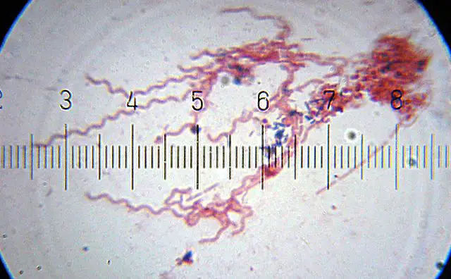
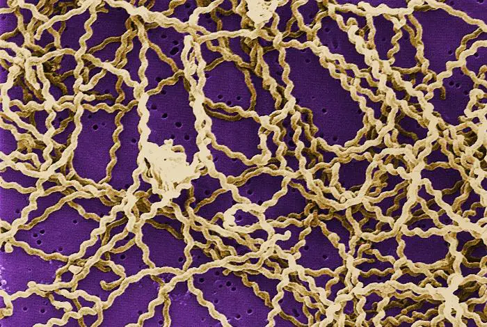
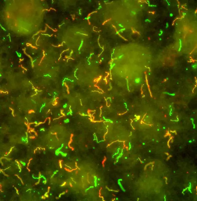
Spirochetes (spirochaetes) are Gram-negative, spiral-shaped bacteria of the phylum Spirochaetes. In nature, they may exist as free-living bacteria, symbionts, or as parasites capable of causing diseases in animals. As such, they are widely distributed in nature, exhibiting varying characteristics in shape and size etc. However, they are all characterized by a distinctive diderm (double-membrane).
Some members of this group include:
- Treponema
- Spironema
- Leptospira
- Spirochaeta
- Borrelia
Classification of Spirochetes
Domain: Bacteria - A large group of single-celled prokaryotes (lacking membrane-bound organelles) found in just about any environment across the world (soil, water, air, hot streams etc). They may exist as parasites, free-living organisms or as symbionts with some of the species being very beneficial to man.
Phylum: Spirochaetes - Bacterial cells characterized by a unique diderm (double-membrane) that gives them their Gram-negative characteristic. They are generally thin with a spiral-shaped appearance and possess flagella commonly known as axial filaments.
Order: Spirochaetales - Helically-shaped bacteria capable of locomotion. They display significant variation in their physiology and distribution. In nature, they may exist as facultative anaerobes, obligate anaerobes, or obligate aerobes.
This order is further divided into three major phylogenetic families including; Spirochaetaceae, Brachyspiraceae, and Leptospiraceae.
Ecology and Distribution
As a group of bacteria, spirochetes are widely distributed in nature and can be found in different environments across the world. Many of the species have been shown to exist as free-living organisms and can be found in various habitats in water (surface water/freshwater), lakes, salt marsh sediments, mud, sediments, and deep-sea vents among others.
Apart from the species found in these habitats, others form an association with various hosts (termites, protozoa, mammals, etc) and are therefore found living within these hosts (in the intestine). While some of the species are beneficial, some are pathogenic and tend to cause diseases (e.g. Lyme disease, dysentery, etc).
Because of the diversity between species and where they are found in nature, spirochetes have also been classified based on their distribution. Obligatory aerobic spirochetes (need oxygen for metabolism) can be found in water and soil as free-living organisms with some species living as pathogens in their hosts.
Members of the genus Leptospira are classified as obligatory aerobic spirochetes. Anaerobic and facultative anaerobic spirochetes exist as free-living forms and include members of the genus Spirochaeta. They are mostly found in different environments where they survive on a variety of organic matter (disaccharides, pentoses, and hexoses, etc).
Some of the other habitats in which spirochetes can be found include:
- High salinity ponds
- Hot/boiling water springs
Characteristics of Spirochetes
Cell structure and Morphology
As the name suggests, spirochetes have a spiral morphology that has been used to classify them based on morphology. From early records, it's believed that spirochetes were first observed under the microscope by Van Leeuwenhoek, who noted that in their motion, they "bent their body into curves in going forwards" (Holt, 1978, p. 115).
Today, it's well understood that this morphology is a result of the flexible peptidoglycan cell wall wound by several axial fibrils.
Along with the cell wall, these fibrils are in turn covered by an outer membrane similar to the membrane found in Gram-negative bacteria. The axial fibril, which has a similar structure to flagellum found in bacteria, consists of a shaft and a covering sheath. It also consists of an insertion apparatus that differentiate into a terminal knob.
Depending on the species, the shaft has been shown to either have a filamentous or globular substructure. While it has similarities to the shaft found in bacterial flagella, it's located between the inner and outer membranes (within the periplasmic space) along the length of the organism.
As already mentioned, the shaft (filament) of the axil fibril is covered by a sheath (covering that encloses the shaft). When the sheath is removed, the internal core is estimated to range between 10 and 16nm in diameter. Apart from giving the organism its shape, the axial fibrils (axial filaments) have been shown to contribute to the movement of spirochetes through a twisting motion.
* The axial filaments/fibrils are also commonly referred to as the endocellular flagella in spirochetes.
Cell Body
The protoplasmic cylinder is located beneath the outer sheath. This is the cell body and consists of a cell wall, cytoplasmic membrane as well as cell contents (cytoplasmic contents) enclosed within. Like other bacteria, the internal environment of the cell has a simple arrangement consisting of a nucleoid, mesosomes, vacuoles, and ribosome.
* The cell-wall-cell membrane complex is referred to as parietocytoplasmic membrane.
Outer Structural Features
Like a number of other bacteria, the cell body of spirochetes is enclosed within several layers. These include the outer and inner membrane, the peptidoglycan layer as well as the cytoplasmic membrane.
Some of the species (e.g. Treponema pallidum) have been shown to contain a slime layer covering the cell. In these species, the layer has been associated with serological non-reactivity.
All species, however, have an outer membrane/envelope that surrounds the entire cell. This is particularly important for the survival of the cell given that damage to this membrane results in a loss of intracellular components and the consequent death of the cell.
This membrane is particularly fragile to various adverse conditions during given stages of cell development. When exposed to hypertonic conditions, the outer membrane/envelope of Leptospira bacteria has been shown to separate from the protoplasmic cylinder producing a spherical shape.
Here, it's worth noting that unlike other cell membranes, the outer membrane does not contain a phospholipid bilayer. According to a number of studies, lipids make up about 20 percent of the total dry weight. This layer also consists of a number of proteins including lipoproteins and B-barrel proteins.
Spirochete Peptidoglycan
The peptidoglycan has been shown to make up about 1 percent of the total cell dry weight and has a similar composition to the peptidoglycan found in gram-negative bacteria. While the axil filaments are responsible for the general spiral shape of spirochetes, the peptidoglycan layer has been shown to help maintain this shape. This layer consists of units of disaccharide N-acetyl glucosamine-N-actyl muramic acid that cross-links with pentapeptide chains.
Some of the other components of this peptidoglycan in spirochetes include:
- Glutamic acid
- Muramic acid
- L-ornithine
* Based on a number of studies, it has been demonstrated that in spirochetes, the peptidoglycan layer is associated with the inner membrane rather than the outer sheath.
Spirochetes Metabolism
In spirochetes, metabolism is largely dependent on their distribution and habitat.
Free-Living Spirochetes
The majority of free-living spirochetes are found in the genus Spirochaeta. They can be found in various habitats including freshwater and seawater mud environments that contain hydrogen sulfide. A good example of free-living spirochete is the Spirochaeta isovalerica.
Along with S. litoralis, Spirochaeta isovalerica are obligate anaerobes that survive by fermenting carbohydrates to produce acetate, ethanol, carbon dioxide and hydrogen. Free-living species can also be found in such genus as Leptospira and live in soil and various aquatic environments.
Host-Associated Spirochetes (non-pathogenic)
Through their association with hosts, some spirochetes become pathogenic and cause various diseases. However, there are many non-pathogenic species that co-exist peacefully with their hosts. Good examples of these are the spirochetes found in the intestine of termites where they digest wood material.
On the other hand, Cristispira, found in the digestive tract of various marine and freshwater hosts (e.g. mollusks) contributes to the digestion process. In turn, they are protected from the external environment where they can only survive for a short period of time.
This relationship has also been observed among nitrogen-fixing free-living spirochetes. These spirochetes are commonly found in the hind-gut of termites (wood-feeding termites). In these hosts, spirochetes have been shown to supply as much as 60 percent of the nitrogen in termite biomass.
Pathogenic Spirochetes
Unlike symbiotic spirochetes that live as commensals in their association with the host without causing any harm, some spirochetes cause harm to the host through this association. Treponemes which cause human treponematoses are a good example of pathogenic spirochetes.
Given that they cause disease in human beings, they are distributed in different parts of the world where they can be spread through sexual contact. Some of the other diseases caused by spirochetes include Lyme diseases, yaws, and relapsing fever.
Reproduction
Like a number of other bacteria, spirochetes produce through binary fission. In spirochetes, this process occurs through asexual transverse binary fission. For this to take place, DNA material is first copied. Here, the process is carried out by replication enzymes starting at the origin of replication.
As the DNA is copied, the cell also increases in size. The new chromosomes then move to opposite poles of the cell. This is followed by division of the cytoplasm as a septum begins to form in the middle.
A new dividing wall then starts to form in the middle allowing each cell to have its own cell wall when the two cells divide into two. This means of reproduction produces two similar daughter cells that develop and repeat the process.
Culture Technique used for Borrelia Species
Borrelia is a genus of the phylum spirochete that consists of species responsible for Lyme disease (Lyme borreliosis).
To culture the bacteria, the following are required:
- Blood sample (this can be collected from a patient with Lyme disease)
- Polypropylene Falcon tube
- Red top Vacutainer tube without any additive
- Vacutainer tube that contains EDTA
- Oven/incubator
Procedure
Collect 9.0ml of peripheral blood into three types of tubes include; Falcon tube with 5ml BSK-H, 10.0ml red top Vacutainer tube without any additive and a Vacutainer tube that contains EDTA - In total, there should be three samples collected in three different tubes
Allow to stand for several hours at room temperature - This step allows for serum separation
For each of sample, divide the serum into eight starter cultures
Mix the samples with modified BSK-H media - Here, 15ml glass tubes and 2ml cryotubes can be used to mix the contents
Incubate the samples at 34º C, 5 percent carbon dioxide, and 100 percent humidity - Close the lids loosely to allow for limited gas exchange
After 6 days, record your results
* Other spirochetes species can be obtained from the appropriate host or the environment in which the species is found. Some symbionts can be found in the gut of termites while free-living species can be obtained from aquatic and terrestrial environments etc.
* Given that different species have different characteristics, appropriate media has to be used to culture-specific species.
Gram Stain
While spirochetes are generally regarded as Gram negative bacteria, because they have a thin peptidoglycan, Gram staining is not commonly used to stain them. In the process of staining Borrelia burgdorferi, they have been shown to weakly stain Gram-negative. However, this is by default given that the secondary dye is used last.
Even though they stain when gram stain is used, they are still too small to be seen under the light microscope. For this reason, they can be viewed using a darkfield microscope or stained using such stains like Vincent's stain. This has particularly proven useful for staining Borrelia vincentii.
To try and stain Spirochetes using Gram stain, the following procedure can be used:
· Using a dropper or pipette, place a drop of the sample on a clean glass slide and heat gently in order to fix
· Flood the slide with 0.5percent crystal violet and let it stand for 30 seconds
· Tilt the slide at an angle and gently rinse with water
· Flood the slide with Lugol's iodine
· Cover the sample with iodine solution and allow it to stand for about 3 seconds
· Tilt the slide at an angle and wash with water
· Decolorize using 95 percent ethanol or acetone
· Rinse the slide with water and flood with the counterstain (0.1 percent counterstain)
To get a clear view of spirochetes under a microscope, the following procedure can be used:
Requirements
- Compound microscope
- Clean glass slide
- Pipette
- Cover slip
- Methyl cellulose
- Eppendorf tube
Procedure
· Using a Pasteur pipette, collect water from a temporary puddle so that you obtain contents at the surface
· Place the water sample in an Eppendorf tube
· You can add some methyl cellulose to the sample in order to reduce the rate of motion of the organism
· Using a pipette, place a small drop of the mixture onto a clean glass slide and then cover using a cover slip
· Observe the slide under the microscope at ✕1000
Observation
Under the microscope, the sample will show a number of contents including diatoms and even flagellates. Here, it's possible to see the spirochete moving from one point to another in the field of view. However, spirochetes will be very small in shape especially when compared to the other organisms visible in the field of view.
Using a rich animal system (consisting of other organisms) is particularly useful given that it makes it possible to differentiate the organisms based on size and movement. Spirochetes will appear to be moving in a wave-like manner which can be very rapid if the cellulose was not added.
Return to Bacteria - size, shape and arrangement main page
Return from Spirochetes to MicroscopeMaster home
References
Ali Karami, Meysam Sarshar, Reza Ranjbar, and Rahim Sorouri Zanjani. (2014). The Phylum Spirochaetaceae.
Eva Sapi1, Namrata Pabbati1, Akshita Datar, Ellen M Davies, Amy Rattelle1, and Bruce A Kuo. (2013). Improved Culture Conditions for the Growth and
Detection of Borrelia from Human Serum.
Kenneth J. Ryan and C. George Ray. (2009). Sherris Medical Microbiology, 6th Edition.
Stanley C. Holt. (1978). Anatomy and Chemistry of Spirochetes.
Links
https://www.asmscience.org/content/education/imagegallery/image.3199
http://textbookofbacteriology.net/Lyme.html
Find out how to advertise on MicroscopeMaster!


