Sugar Crystals under the Microscope
Preparation and Observations
Introduction
Crystallization is the process of formation of solid sugar crystals from a sugar solution and it offers a particularly fascinating and rewarding experience for the beginner microscope enthusiast, and there is basically no limit to the scope for exploration, even with relatively simple apparatus.
In this post, we are going to observe the appearance of sugar crystals under the microscope. To get the best of this exploration, the compound microscope and the stereomicroscopes are used but the stereomicroscope offers the best result because of the stereoscopic or 3-Dimensional appearance of the sugar crystal lattice.
Sugar which is the sweet tasting white gritty stuff we all know is sucrose, with 12 carbon atoms, 22 atoms of hydrogen and 11 of the oxygen molecule with the molecular formulae (C12H22O11). Just like every other compound made from a combination of the three chemical elements listed, sucrose is a carbohydrate, a two-sugar carbohydrate (disaccharide).
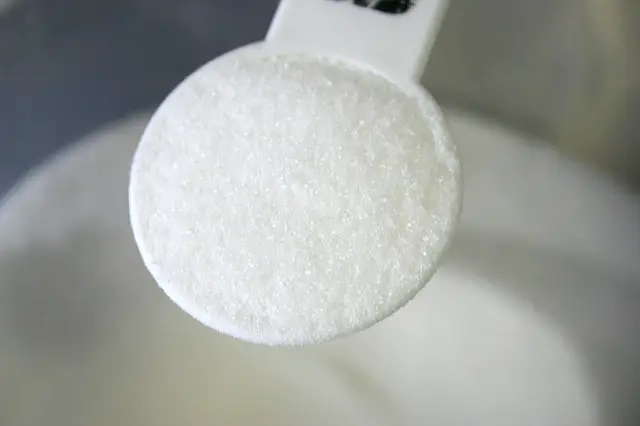
Naturally, sucrose or sugar can be found in a good number of plant sources with the majority being in sugarcane. For this simple experiment, it's best to use granulated sugar which has a grain size of about 0.5 mm across and is mostly used as dietary sugar in coffee or tea.
Demerara sugar can be seen to be crystalline with the unaided eye while granulated sugar is clearly seen to be crystalline when viewed with the hand magnifier and better with a microscope.
The crystalline structure of the caster sugar can only be seen with the compound or other microscopes. Also, it will not be easy to observe the very fine crystalline nature of icing sugar with a microscope, but it depends on the type of microscope used.
Using a Compound Microscope
Required Materials:
- Granulated sugar crystals.
- Microscope slide.
- Compound microscope with power supply and illuminants.
- Spatula
Procedure
1. Turn the revolving turret of the microscope so that the lowest power objective lens is clicked into the 40x position.
2. Put a half-teaspoon of granulated sugar on the microscope slide and hold it in place with the clips.
3. Look through the microscope’s eyepiece and then move the focus knob carefully for the image (sugar crystals) to come into clear focus.
4. Slightly adjust the microscope’s condenser and amount of illumination for optimum light intensity.
5. With the focus knob, carefully place the image into clear focus and also readjust the condenser and amount of illumination for a clear image.
6. Once the image of the sugar sample comes into clear focus with the x10 power objective, you can then switch to the net higher or lower objective to zoom in or out of the image for clarity. You can at this point use a spatula to turn the crystal into different planes for better observation of the crystal geometry.
7. When you’re done with the viewing, lower the stage, then click the objective into the low lens power and take out the slide.
Using the Stereo Microscope
Required Materials:
- Granulated sugar crystals.
- Microscope slide.
- Stereo microscope
- Spatula.
Procedure
1. Mount a half-teaspoon of granulated sugar on the microscope slide on the stage and turn the illumination on.
2. View through the eyepieces and adjust the focus knob until the sugar crystal sample image comes into clear focus.
3. Make some slight adjustments in the distance between the eyepieces. It may be helpful to know your inter-pupillary distance (IPD) before viewing through the eyepiece. This is to enable you to have a simultaneous binocular vision of the sample. With this in place, you will see the crystal in 3-Dimension which has some advantage over the compound microscope as we can see in the observation.
Sugar Crystals Observations
The basic geometry of sugar crystals appear irrespective of the size of the crystal and the crystals do not just appear individually or in isolation but are sometimes like a fused mass or joined with other crystal molecules.
As you can see if you’re following this, they appear like hexagonal pillars that have fallen over at one side and somehow flat on the surface. This crystal appearance makes sense since sugar naturally forms molecules in a monoclinic form. Some crystals are much smaller than others and it might be actually how long they had to grow or the different conditions under which they’ve had to form.
The edges are quite rough and not exactly a perfect hexagon. Since they are 3-Dimensional, with a compound microscope, you will see a fuzzy outline on the edge where there is an out-of-focus section.
For better viewing under the compound microscope, you can either focus on the top of the crystal or you can focus further down its geometry. Therefore, if you go through the full range, you can see the image plane of different areas on the top of the crystal.
This is not the case with the stereomicroscope which is adapted for better viewing of 3-Dimensional crystal lattices. Once the magnification is set correctly, every part of the crystal comes into focus at once and the depth or outline can be observed more clearly.
The crystals look a bit more transparent than they appear on normal unaided view, that’s with just your eyes. You can see the black background behind, blending through the crystals very easily and the crystals almost look like frosted glass or perhaps like an ice that’s out of an ice tray.
Increasing the magnification with the compound microscope to x40, on a backlit setup other than the initial front lit setup, and focusing on the top of the crystal, you can use the spatula to shift the position and plane of the crystal around.
Focusing on the top, you can see the crystals have a fairly flat surface with the corners quite chopped off. Actually, they appear to be rounded off quite a lot and the surfaces seem to be worn down as well. This may be because of the continued impact of the crystals against one another causing the erosion of some of the edges.
Conclusion
The appearance of sugar crystals under the microscope both under the compound microscope and stereo-microscope is an enjoyable experiment.
With the stereo-microscope, there’s more in-depth focusing of the entire crystal structure as the microscope is better adapted for 3-dimensional viewing otherwise known as depth perception. The crystals appear like hexagonal pillars that have fallen over at one side and somehow flat on the surface.
This crystal appearance makes sense since sugar naturally
forms molecules in a monoclinic form. Some crystals are much smaller than
others and may be due to how long they had to grow or the different
conditions that may have affected the process of crystallization.
Check out other easy microscope experiments: Onion Cells, Leaves, Hair, Spider's Web, Cheek Cells and Cork Cells under the Microscope
Return from Sugar Crystals under the Microscope to Microscope Experiments Main Page
Return to MicroscopeMaster Home
References
Urbano, L., 2011. Salt and Sugar Under the Microscope, Retrieved May 9th, 2018, from Montessori Muddle: http://MontessoriMuddle.org/
Find out how to advertise on MicroscopeMaster!

![Sugar Crystals by Edal Anton Lefterov [CC BY-SA 3.0 (https://creativecommons.org/licenses/by-sa/3.0) or GFDL (http://www.gnu.org/copyleft/fdl.html)], from Wikimedia Commons Sugar Crystals by Edal Anton Lefterov [CC BY-SA 3.0 (https://creativecommons.org/licenses/by-sa/3.0) or GFDL (http://www.gnu.org/copyleft/fdl.html)], from Wikimedia Commons](https://www.microscopemaster.com/images/512px-Sugar-Crystals-magnified.jpg)


