Coelomates, Acoelomates, and Pseudocoelomates
** Differences and Examples
Overview
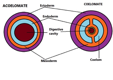
Located between the alimentary canal and body wall, the coelom is the principal body cavity found in many animals.
The presence or absence of this fluid-filled cavity is one of the factors used to classify animals. Animals in which the coelom is lined by the mesoderm are known as coelomates while those in which the cavity is absent are called acoelomates.
In some metazoans, the body cavity is not entirely lined by mesoderm (the endothelial lining found in coelomates). For this reason, they are referred to as pseudocoelomates (do not have a true coelom).
Examples of acoelomates:
- Fasciola hepatica
- Taenia saginata
- Schistosoma japonicum
Examples of pseudocoelomates:
- Ascaris lumbricoides
- Priapulus caudatus
- Trichinella spiralis
Examples of coelomates:
- Human beings
- Elephants
- Birds
- Fish
Characteristics of Acoelomates
Members of the phylum Platyhelminthes (flatworms) are some of the most popular acoelomates. Other phyla in this group include Gastrotricha and Nemertea.
Like many other animals, they are triploblastic organisms and thus have a body structure that is derived from three embryonic germ layers; ectoderm, mesoderm, and endoderm.
Whereas ectoderm gives rise to the epidermis, the gastrodermis is derived from the endoderm layer. The mesoderm, on the other hand, gives rise to the muscle fibers, mesenchyme, and specific organs.
In many other animals, the mesoderm also forms two sheets of tissue that separate in order to form the coelom. As this body cavity forms, it is lined with mesodermal tissue and occupies the space between the intestinal canal and the body wall.
In Platyhelminthes, however, this space is filled with mesenchymal tissue. Because they do not have a respiratory system nor an enclosed circulatory system, the simple, incomplete digestive system is the only body cavity they have (not a true coelom).
Acoelomate mesenchyme
The mesenchyme consists of several cells types that include:
Myocytes - Myocytes make up most of the cells found in the mesenchyme and are the primary component of formed muscles. The mesenchyme also contains undifferentiated myoblasts within the regeneration blastemas.
Generally, differentiated myocytes have two main parts which include the contractile myofibrils and the nucleated myocyton (non-contractile part of the cell body).
In triclad turbellarians (members of the order Tricladida) the cytoplasmic region joining the two parts has been shown to be relatively broad. This has been observed in a number of other species including Oochoristica anolis and G. urna among others (the region is thinner in species like Mesocestoides lineatus and Hymenolepis diminuta).
The degree to which the two parts are separated varies from one group of species to another.
Though they do not contract, myocytons serve a number of important functions and are thus divided into several categories. These include secretory myocytons (involved in production of proteins), storage myocytons, and secretory/storage myocytons (store glycogen and lipids).
Neoblasts - Neoblasts are mesenchymal, generative cells found in Platyhelminthes. They have been shown to play an important role in wound healing, organ replacement under certain conditions, and asexual reproduction.
Although they can be found in some acoelomate forms (e.g. turbellarians and juvenile cestodes), they are absent in others (e.g. adult cestodes and digeneans).
* Some of the other cells that can be found in the mesenchyme include vacuolated chordoid cells, granular, calcareous corpuscle cells, pigmented cells, and fixed parenchymal cells.
* Mesenchymal gland cells found in turbellarians are involved in the production of a slimy substance that contributes to locomotion as well as the secretion of an adhesive material used to capture prey.
Some of the other characteristics of acoelomates include:
- An organ-system organization
- A simple nervous system
- Exhibit bilateral symmetry
Coelomates
As the name suggests, coelomates are organisms that have a coelom (body cavity).
For an organism to qualify as a coelomate, it must have a true coelom (where the body cavity is fully lined with the mesodermal epithelium).
Like acoelomates, coelomates are triploblastic and thus develop from embryo with three germ layers. However, they are distinguished from acoelomates by the fact that they have a body cavity (fully lined with the mesodermal epithelium).
Some examples of coelomates include:
- Slugs
- Earthworms
- Snails
- Arthropods
- Mammals
Coelomates are divided into two main categories that include:
- Protostomes
- Deuterostomes
Protostomes (e.g. mollusks) - The name protostome is derived from the Greek words "Proto" meaning first and "Stoma" which means mouth.
In protostomes, the blastopore first gives rise to the mouth during embryonic development. Typically, this is caused by the fusion of the lateral lips. In many coelomates, the blastopore also gives rise to the anus as development continues (the mouth is formed first).
* The blastopore is the first opening (invagination) during gastrulation.
Deuterostome (e.g. echinoderms) - In deuterostomes, the blastopore gives rise to the anus.
* Although coelomates are divided into two categories (deuterostome and protostome), it's worth noting that several changes have been observed in the development of protostomes in various animals. For this reason, this type of classification has faced some opposition.
Coelom Formation
Following fertilization, the zygote undergoes cellular divisions known through a process called cleavage. This results in the formation of a solid mass of cells (blastomeres) known as morula and eventually blastula (a hollow sphere of blastomeres).
As the process continues, a central cavity (blastocoel) is formed in the blastula. Cells at one end then invaginate to form the gastrula. The gastrula contains an open cavity (lined by endoderm) known as the archenteron and an opening known as a blastopore.
At this stage, the embryo has two layers of cells, the ectoderm and the endoderm. The blastocoel is located between them. As the mesoderm develops between the two layers, it lines the cavity forming a true coelom.
In deuterostomes, the coelom is formed through a process known as enterocoelous.
Also known as enterocoely, this process involves the formation of coelom from pouches of the archenteron during gastrulation. As the pouches develop and separate from the archenteron, the cavity formed becomes the coelom.
The mesoderm continues to develop as the inner wall (lining the alimentary canal) and outer wall (along the body wall) continue to grow in size and ultimately extend towards each other.
When they meet and fuse, they completely separate the endoderm and ectoderm.
Schizocoelous - Schizocoelous is the type of coelom formation that occurs in protostomes.
Also known as schizocoely, this type of coelom formation is characterized by the splitting of mesodermal embryonic tissue. This type of cavity is commonly referred to as a schizocoel.
In coelomates, the coelom serves a number of important functions that include:
Shock absorption - The coelom is not an empty space. Rather, it's filled with a fluid known as coelomic fluid. By separating the inner organs of the upper body from the outer frame, this fluid-filled cavity helps protect these organs from trauma and mechanical shock.
Organ growth - The coelom also creates the space that organs need to grow. As mentioned, this space is filled with coelomic fluid. Unlike rigid frames, the fluid allows organs like the digestive tract room to grow and increase in size. This is also important during pregnancy.
Hydrostatic skeleton - Invertebrates like earthworms do not have an inner skeleton or exoskeleton: In this case, the coelom serves as a hydrostatic skeleton that not only maintains the general shape but also promotes movement.
Exchange of material and excretion of waste productions - Coelomic fluid within the coelom plays an important role in the movement of various materials (nutrients and gases as well as waste products) to various parts of the body.
For instance, when nutrients are absorbed into the fluid; they can be transported to various parts of the body that require them. As well, waste products can be released into the coelomic fluid before they are completely removed from the body.
* In invertebrates, cells known as coelomocytes (macrophage-like cells) are involved in a number of immune activities including phagocytosis and the production of humoral factors.
Pseudocoelomates
Although pseudocoelomates, like coelomates, have a coelom, the coelom is located between the mesoderm and endoderm. For this reason, it's not considered a true coelom.
Some examples of pseudocoelomates include members of the phyla Nematoda and Rotifera.
In most pseudocoelomates, the Pseudocoelom is formed later in embryonic development. The fluid contents that fill the space originate from blastocoel, the fluid associated with the early blastomeres.
Once it's fully developed, this cavity is particularly larger along the mid-body region but declines near the head and the tail tip of the organism. Because the cavity is partially lined by the endoderm, it's closer to various body organs.
Here, it bathes the tissues and also performs a variety of functions usually performed by the circulatory and respiratory systems.
The fluid has been shown to play a role in establishing ionic equilibrium by balancing the osmotic content of the tissues. As well, it acts as a hydrostatic skeleton by counteracting the forces of the muscles.
* The coelom in pseudocoelomates is known as pseudocoel or pseudocoelom.
Like the other animals (coelomates and acoelomates), pseudocoelomates are triploblastic organisms and exhibit bilateral symmetry.
The majority of species in this group have a vermiform body morphology (worm-like). With the exception of a few species (e.g. acanthocephalans), the majority have a complete digestive system but no circulatory or respiratory system.
See also:
Learning about Kingdom Animalia
Return to phylum Platyhelminthes
Return from Acoelomate, Pseudocoelomate, and Coelomate" to MicroscopeMaster home
References
Conn, D. B. (1993). The Biology of Flatworms (Platyhelminthes): Parenchyma Cells and Extracellular
Matrices. Transactions of the American Microscopical Society, Vol. 112, No. 4. (Oct., 1993), pp. 241-261.
Giribert, G. (2014). Protostomes: The Greatest Animal Diversity. Sinauer Associates
Rieger, R. and Purschke, G. (2005). The coelom and the origin of the annelid body plan. Part of the Developments in Hydrobiology book series.
Links
https://jan.ucc.nau.edu/allred/bio190/pseudocoelomate/lesson.html
https://www.wormatlas.org/hermaphrodite/pericellular/mainframe.htm
Find out how to advertise on MicroscopeMaster!


