Phylum Percolozoa
** Examples and Characteristics
Overview
Phylum Percolozoa is within the kingdom Eukaryota consisting of colorless protozoa. They can be found in aquatic and terrestrial environments and have been shown to change form. In these environments, the majority of species are free-living with Naegleria fowleri as the only pathogen in humans.
Currently, members of this group are divided into four main classes that include:
- Pharyngomonadea
- Percolatea
- Lyromonadea
- Heterolobosea
Phylum Percolozoa: Characteristics of Class Pharyngomonadea
The class Pharyngomonadea in phylum Percolozoa consists of a single genus known as Pharyngomonas within the family Pharyngomonadidae.
Morphology
Based on studies involving Pharyngomonas kirbyi, members of this group were shown to go through a life cycle comprising three stages that include flagellate, amoeboid, and cyst.
For this reason, there are a number of variations depending on the particular stage of life:
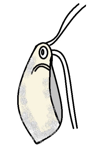
Flagellates - The flagellated cells are characterized by two pairs of flagella (posterior and anterior flagella), a cytopharynx located in the ventral groove, longitudinal grooves on the ventral side, while the nucleus is located near the base of the flagella.
Generally, these cells are spindle-shaped and laterally flattened with the posterior part being more attenuated than the posterior. However, in some of the species, the anterior might appear more rounded with a pointed posterior end. Depending on the species, the cells may range from 9 to 19 um in length and 3-8 um in diameter.
* The flagella in this stage might be 1.5 to 2 times the length of the cell.
* While flagella differentiate into posterior and anterior flagella, they originate from the anterior end of the cell. For this reason, the posterior flagella are longer in length.
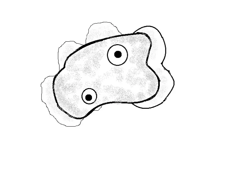
Amoeboid - The amoeboid may be lobosean or heterolobosean. Depending on the species, lobosean forms may be ovoid, flabellate, or rectangular in shape, ranging between 10 to 32 um in length and 8- 28 um in diameter/width. They are characterized by a flattened cell body with a serrated anterior edge as well as a dominant hyaloplasm (part of cytoplasm) that occupies about a third of the cell.
While they lack flagella, they can move by means of filiform pseudopodia that form at the anterior and lateral edges of the cell body. However, long, thin flagella are also produced at the posterior end.
Compared to lobosean forms, heterolobosean forms are rare and resemble amoeboid forms of Heterolobosea (to be discussed later). However, they also move by means of filiform pseudopodia and also produce eruptive pseudopodia at the anterior and lateral edges of the cell.
Cysts- Cysts are generally spherical or ovoid in shape and consist of a thick wall. The thick wall serves as a protective layer that protects them from fluctuations in salinity in the environment.
Depending on the species, they vary in size from 9 to 18 um in diameter. When conditions improve (availability of food material at 5 to 10°C with the right saline concentration), the cyst gives rise to the amoeboid form which can then transform into a flagellate form.
Habitat
Members of this group can be found living freely in aquatic environments including lakes and some marine habitats. For the most part, they prefer environments with salinity above 15% and below 300%.
Nutrition
In their natural habitats, members of the class Pharyngomonadea in phylum Percolozoa are predators of bacteria. In marine environments, for instance, the species Pharyngomonas kirbyi uses the posterior flagella to not only swim but also direct the prey to the central groove before reaching the cytopharynx.
* Reproduction is achieved through binary fission.
Some of the species within the class Pharyngomonadea include:
- Pharyngomonas turkanaensis
- Pharyngomonas kirbyi
- Macropharyngomonas halophila
Phylum Percolozoa: Characteristics of Class Heterolobosea
Members of the class Heterolobosea in phylum Percolozoa are some of the most common species within the Phylum Percolozoa. Currently, the group is estimated to consist of over 150 species that can be found in different habitats across the globe.
Morphology and Life Cycle
Like Percolatea, the majority of Heterolobosea species are unicellular organisms that go through three life stages consisting of the amoeboid, flagellate, and cyst (the cyst is the resting stage).
A few of the species (e.g. Stephanopogon, G. flavescens, and Willaertia magna, etc.) have been shown to be multinucleate but the majority are uninucleate.
* Amoeboflagellates include species with three stages in their lifecycle.
Amoeboid stage - The amoeboid forms (amoebae) are characterized by a lobose morphology and thus possess broad cylindrical pseudopodia. Rather than forming pseudopodial extension, the cell behaves like a single large pseudopodium with a wormlike movement.
For this reason, they are also referred to as monopodial forms (or limax type). Here, motility is made possible by eruptive lobopodia that allows for rapid movements. While the majority of species are monopodial, a few species (e.g. Fumarolamoeba ceborucoi) are capable of forming short subpseudopodia that radiate from the cell body that enhances motility.
The majority of species have also been shown to form bulb-like excretory organs known as uroid (uroidal filaments) at the posterior end. Waste material from the cell body accumulates in the organ for a given period of time before they are released into the environment.
Some of the other morphological traits of amoeboid forms include:
- Lack cytoskeleton-underlain cytostomes.
- Naegleria fowleri, which can cause disease in human beings uses amoebastomes for phagocytosis
Flagellates - For many of the species, the amoeboid form can transform into a flagellate under stressful environmental conditions like changes in temperature or fluctuations in food sources. However, the amoeba can also transform into cysts when conditions drastically decline.
The majority of flagellates possess a subapical cytostome for feeding. In some of the species, however, this structure opens at the anterior part of the cell. As the name suggests, flagellates use flagella for motility.
Depending on the species, there are two (e.g members of the genera Euplaesiobystra and Pocheina, etc.) or four flagella (e.g. members of the genera Tetramastigamoeba and Willaertia, etc.) that are parallel to each other. Here, flagella originate from the anterior part of the cell.
While the number of flagella varies between members of different genera, some studies have also identified variations among members of the same species (e.g. Tetramitus jugosus).
* The number of flagella varies between two and more than four for the species Tetramitus jugosus.
* Posterior flagella beat slowly compared to the anterior flagella.
* Some species (e.g. members of the genus Stephanopogon) have over 100 flagella.
While the flagella originate from the anterior part of the cell body, they may vary in length. For instance, among members of the species Percolomonas, some of the flagella are longer than others. However, they all beat simultaneously to create a current that moves food particles towards the cytostome.
For some of the species, food material may attach to the substrate before being moved to the cytostome for consumption. Apart from serving an important role in the feeding process, flagella also promote motility, allowing the organism to swim in the water.
Some of the other characteristics of flagellates in this group include:
- Some species have a feeding groove while others use elongated cytostome for feeding
- Some of the species (e.g. members of the genus Tetramitus) are characterized by rim-like structures at the anterior part of the cell body
- The basal bodies of some species may be organized in an arrangement known as double bikont
- The cell body might be vase-shaped, curved, or elongated
- The cytostome which is associated with the ventral barbs is slit-shaped
* Some species do not go through the flagellate stage. These include members of the genus Vahlkamfia.
Cyst forms - As mentioned, amoeboid forms can transform into cysts in the event of poor environmental conditions. These forms are characterized by two main layers namely, the ectocyst (outermost layer) and endocyst (inner layer).
Depending on the species, the outer surface of the cyst may appear wrinkled, rough, or smooth in texture. In some of the species (e.g. members of the genera Pernina, Willaertia, and Naegleria), the cysts have one or multiple pores that contribute to excysting under conducive conditions. For the majority of species, pores are absent and the external wall has to rupture for the cell to give rise to other forms.
* In some species (e.g. A. helenhemmesae), the life cycle has been shown to consist of an additional stage involving a sorocarp which is a multicellular fruiting body. These fruiting bodies may vary in shape from one species to another.
Habitat
Members of the class Heterolobosea can be found in a variety of habitats across the globe where they exist as heterotrophs. The majority of species can be found in terrestrial and freshwater habitats.
Some species can be found in marine habitats with salinity levels of between 30 and 50% (e.g. some members of the genera Stephanopogon and Pseudovahlkampfia). In these environments, they survive by feeding on bacteria (bacteriovores) but have also been shown to feed on diatoms and various eukaryotes.
While many species can be found in freshwater environments, other species have adapted to live in aquatic environments with varying conditions.
These include:
- Hypersaline habitat - E.g. Euplaesiobystra hypersalinica and Tulamoeba peronaphora
- Acidic habitats - E. g Tetramitus thermacidophilus
- Thermophiles - E.g Oramoeba fumarolia
- Psychrophiles - E.g Vahlkampfia signyensis
- Halophiles - E.g. Sawyeria marylandensis
While the majority of heterolobosea species in phylum Percolozoa exist as free-living organisms in various habitats across the world, a few species exist as endosymbionts or as pathogens. As such, they can be found in association with other organisms (host).
Naegleria fowleri is one of the most common pathogens of human and animal hosts. In these hosts, the organism is responsible for meningoencephalitis which can be fatal.
Naegleria fowleri can gain entry into the body of the host through the nasal chamber (e.g. during swimming in freshwater) from where it can migrate to the olfactory neuroepithelium to reach the central nervous system. Infection of the cells of the CNS can result in death within 14 days if proper care is not provided.
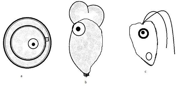
Diagrammatic representation of the three life stages of Naegleria species in phylum Percolozoa:
A – Cyst
B – Amoeboid
C – Flagellate
Phylum Percolozoa: Characteristics of Class Lyromonadea
The class Lyromonadea in phylum Percolozoa consists of a single-family (Lyromonadidae) and four genera namely, Lyromonas, Sawyeria, Monopylocytis, and Psalteriomonas.
* Before the concept of classification that was introduced by Cavalier-Smith and Nikolaev in 2008, these organisms were all grouped within the class Heterolobosea.
Genus Psalteriomonas
The genus Psalteriomonas consists of a few species with Psalteriomonas lanterna and Psalteriomonas vulgaris being some of the most popular members.
Based on a number of studies, these species have been shown to be microaerophilic amoeboflagellates and thus capable of surviving in areas with little oxygen concentration. As amoeboflagellates, they can transform between the flagellate and amoeboid stages.
Compared to the flagellate, amoeboid forms lack flagella and move by means of pseudopodia. However, both forms have a large globule located at the center of the cell body. In flagellates, the flagella (four in most species) are located at the posterior end of the cell and serve an important role in motility and feeding.
They are also characterized by modified mitochondria with surrounding cisterns and rough E.R.
Members of this genus are commonly found in freshwater sediments where they exist as free-living organisms. Here, some species have been shown to form a symbiotic relationship with chemoautotrophic bacteria (methanogenic bacteria) while others survive by feeding on bacteria.
Genus Lyromonas
The genus Lyromonas consists of a single species, Lyromonas vulgaris. It's an anaerobic flagellate characterized by four flagella located at the anterior part of the cell body. The cell also consists of a longitudinal ventral groove, a kinetid, and globule.
It can be found in freshwater anoxic sediments in association with methanobacterium formicium (endosymbiont) or feeding on other bacteria.
Genus Sawyeria
The genus Sawyeria also consists of a single species, Sawyeria marylandensis, which can be found in anaerobic sediments. Like some of the other members in this group, Sawyeria marylandensis lacks typical mitochondria. However, unlike some of the other group members, the species is a gymnamoeba and does not go through the flagellate and cyst forms.
Although it is commonly found in anaerobic sediments, it can grow in environments with little levels of oxygen and thus qualifies as a microaerophile. The amoeboid form is characterized by a single large nucleus and mitochondrion-related organelles (MRO).
Genus Monopylocytis
The genus Monopylocytis consists of a few species include Monopylocytis elegans, Monopylocytis robusta, and Monopylocytis disparata among others.
Members of this group are found in anoxic environments, including freshwater and marine environments where they may exist as flagellates or in amoeboid forms.
Under harsh conditions, some species (e.g. Monopylocystis visvesvarai) can transform into cysts for survival. Flagellate forms are characterized by two pairs of flagella, a harp-like structure along the mastigont, and a ventral groove.
Phylum Percolozoa: Characteristics of Class Percolatea
Members of the class Percolatea in phylum Percolozoa are confined into two main genera that include Stephanopogon and Percolomonas.
The following are their respective characteristics:
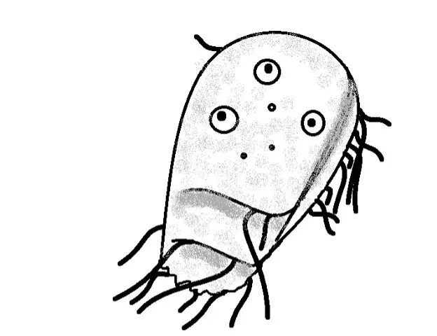
Stephanopogon - Like many other species of the phylum Percolozoa, members of the genus Stephanopogon are unicellular organisms. They only consist of two life stages, flagellate and cyst, without the amoeboid stage. Depending on the species, they vary in size from 20 to 90 um in length.
Some of the species (e.g. S. paramesnili) have been shown to grow about 110 um length in culture. They are characterized by a dorsoventrally flattened shape with 2 or more isomorphic nuclei.
In species like Stephanopogon minuta, the cell body consists of rows of cilia/flagella with three triangular barbs at the anterior end. The rows of flagella (6 to 14 depending on the species) are located at the ventral surface of the cell body.
While cysts are formed due to changes in the environment (e.g. in the absence of food sources), they also play an important role during reproduction. Here, the cytoplasm and nuclei separate into two within the cyst to form two daughter cells.
Following this initial division, the nucleus in each of the daughter cells undergoes a second round of division before the cells excyst and develop into mature flagellates capable of swimming around.
Some of the other morphological characteristics include:
- Have a tapered anterior end
- Right and left sides are curved
- Have a contractile terminal vacuole
* In marine habitats, they feed on a variety of organisms including bacteria and diatoms.
Percolomonas - Members of the genus Percolomonas are also free-living flagellates commonly found in marine environments. Their cell bodies (between 6 and 12 um in length) are characterized by four flagella/cilia (one long cilia and three short ones), a ventral groove, and a pocket-like pharynx.
In marine habitats, different species can be found in areas with varying salinity levels (between 70 and 280 percent salinity).
What does phylum mean in biology?
Return from learning about phylum Percolozoa to MicroscopeMaster home
References
Andrey O. Plotnikov, Alexander Mylnikov, and Elena A Selivanova. (2015). Morphology and life cycle of amoeboflagellate Pharyngomonas sp. (Heterolobosea, Excavata) from hypersaline inland Razval Lake.
Denis V. Tikhonenkov et al. (2019). Ecological and evolutionary patterns in the enigmatic protist genus Percolomonas (Heterolobosea; Discoba) from diverse habitats.
Mahmut Caliskan. (2012). Genetic Diversity in Microorganisms.
Tomáš Pánek and Ivan Čepička. (2012). Diversity of Heterolobosea.
Links
https://www.sciencedirect.com/topics/agricultural-and-biological-sciences/heterolobosea
http://tolweb.org/Heterolobosea/96360
http://tolweb.org/Stephanopogon
Find out how to advertise on MicroscopeMaster!


