What is Cytokinesis? Vs Mitosis
Cytokinesis refers to the division of the cytoplasm during cell division (mitosis). While this is the last event of cell division, it starts early on during the anaphase stage of mitosis in most eukaryotic cells.
Because it involves the separation/division of the cytoplasm, cytokinesis serves to divide the cell into two with each of the daughter cells containing the same cell contents as the parent cell.
In Animal Cells
As mentioned, cytokinesis starts during the anaphase stage of mitosis. This is important for several reasons. Before the metaphase stage, genetic material of the cell is not yet condensed and the nucleus is still intact. However, during metaphase, genetic material first condenses into chromosomes which makes them visible.
This stage of mitosis is also an important one given that it's the stage at which the chromosomes start aligning at the central part of the cell so that they can be divided. Here, it's worth noting that cytokinesis is a tightly coordinated process that follows the separation of chromosomes.
This is important because it ensures that as the cell continues to separate, each of the new daughter cells receives an equal number of chromosomes. For this reason, cytokinesis cannot occur during metaphase and therefore has to wait until the separation of chromosomes start (during anaphase).
The anaphase stage, following metaphase, is characterized by the development of spindle fibers. These are also known as central spindle and consists of an array of microtubules.
Here, centrioles, contained in the centrosome, are responsible for the production of microtubules that form the central fibers. Once they are produced, the array of microtubules extend to different directions within the cytoplasm forming the spindle.
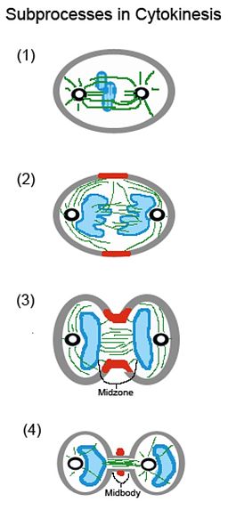
As they extend, some of the microtubules also attach and connect to the chromosomes at kinetochores allowing the separation to begin. As the spindle fibers separate the chromosomes, a cleavage furrow is formed between the chromosomes. This is also the region/point at which the cytoplasm starts dividing.
While some studies attribute the onset of anaphase chromosomes, this is attributed to the activities of the spindle by other studies. Regardless, it's the separation of chromosomes and the consequent creation of a cleavage furrow between the chromosomes that initiates cytoplasm division.
* In plants and yeast, position of the cleavage furrow is said to be independent of the mitotic spindle. Rather, it's the furrow that has been shown to orient the fibers for chromosomal separation to occur. In animal cells, new reports are showing that spindle fibers are responsible for the position of the cleavage furrow.
* In higher plants, microtubule organizing centers (centrosome) is absent. However, studies have shown spindle fibers to originate from a region in the nuclear membrane during the interphase stage.
From here, microtubules that form the fibers radiate to the cell cortex located at the inner part of the cell membrane. This is particularly important because it allows the nucleus to be positioned at the central part of the cell.
In addition, this has been associated with the development of phragmosome which is a cytoplasmic diaphragm that forms in preparation for cell division. In addition to the phragmosome, the microtubules are also involved in the medial rings known as preprophase bands around the phragmosome.
During the early stages of mitosis, these rings narrow to create the place of cell division. While the structure is disassembled before the metaphase stage, the plane created when it narrows marks the site of division which is also the point at which the cytoplasm starts separating.
In animals, the cleavage furrow consists of a contractile ring made up of actin and myosin filaments. Here, it's also worth noting that these are the components of muscle and are involved in muscle contraction.
Accumulation of these components (actin and myosin) is influenced by a number of factors. These include Rho proteins, Aurora B, and Polo proteins. At the cell equator, these factors stimulate the recruitment of formins which in turn results in the nucleation of actin filaments as well as myosin localization.
In addition to actin and myosin (Myosin II), the furrow also consists of two important scaffolding proteins (anillin and septins) that are involved in the localization of the furrow as well as its stability.
During cytokinesis, the contractile ring contracts (undergoes ingression) thus creating an intercellular bridge between the two cells before they completely separate.
Here, studies have shown the contraction of the ring is as a result of the actions of the filaments in the ring. Because of filament sliding, the ring is shortened which in turn produces the force that results in the ingression of the furrow.
Before the two daughter cells separate, the site of division is characterized by a number of events. One of these events is the formation of the midbody. It consists of remnants of interdigitated microtubules as well as interpolar microtubules from the spindle fibers.
Here, they are packed together to form the midbody structure between the two cells. The other common event at this site is the delivery of intercellular vesicles. These vesicles are important in that they introduce new membrane that is used to compensate for the extra surface area.
Originating from the Golgi bodies, these vesicles also bring proteins needed at the furrow. Recycling endosomes and the endocytic pathway are involved in remodeling of the membrane before cytokinesis is completed.
It's the activity of the recycling endosomes and the aforementioned pathway that ultimately splits the membrane resulting in two similar daughter cells. This action is commonly known as abscission.

* Unlike animal cells, plant cells have a cell wall which is tougher than the plasma membrane and thus makes it difficult for the cell to just split into two by pinching and separation of the cell membrane.
For this reason, division of the cell, and thus the cytoplasm, involves the formation of a cell plate (consists of secretory vesicles). As mentioned, a structure known as phragmosome is first formed in preparation for cell division.
During cell division, the phragmoplast which consists of microtubules is involved in division of the nucleus to form two nuclei. Separation of the nuclei is then followed by the formation of the cell plate in the phragmoplast.
The vesicles, which contain a matrix of molecules (in the cell plate), form the plasma membrane on both sides of the plate which allows a new part of the cell membrane to be formed for each of the daughter cells.
This new membrane then fuses with the original part of the membrane for each daughter cell to form a complete membrane for each of the cells. Carbohydrate molecules in the vesicles then form a middle lamella between the two membranes. Cellulose is then secreted by each of the new cells which form the primary cell wall between the cells.

Mitosis Vs Cytokinesis
Mitosis and cytokinesis are both part of cell division. While there are several differences between them, the two overlap during cell division. Generally, mitosis is similar to karyokinesis given that it involves division of the cell nucleus.
The process is divided into four main stages which include prophase, metaphase (often divided into prometaphase and metaphase), anaphase, and telophase.
In each of these stages, the nucleus (chromosomes) undergo important changes that ultimately result in the division of chromosomes into two equal sets. In prophase, condensin is involved in the condensation of the chromosomes.
This is followed by metaphase which is characterized by the alignment of the chromosomes at the cell equator where they are attached to the spindle fiber.
Sister chromatids are separated in anaphase as they move to the opposite poles of the cell. This process ends when the chromosomes have migrated to the opposite poles of the cells and a nuclear membrane starts to form around the new set of chromosomes.
Unlike mitosis, cytokinesis starts during the anaphase stage when the sister chromatids are being separated. This is because the site/region at which the cytoplasm starts separating/dividing is dependent on the separation of chromatids.
Following the separation of the sister chromatids, division of the cytoplasm also serves to divide cell organelles given that they are dispersed in the cytoplasm. This ensures that each of the new daughter cells contains the same cellular components as the parent cell.
While mitosis ends at the telophase stage, cytokinesis does not end until after the end of this stage. As such, cytokinesis is truly the last phase of cell division. As mentioned, the end of cytokinesis is characterized by abscission where the cell membrane is completely separated to two daughter cells.
Therefore, when the two processes overlap at a given point, there are some differences with regards to the main stages in which they occur as well as regarding their respective functions - Mitosis serves to divide the sister chromatids, and thus the chromosomes, while cytokinesis divides the cytoplasm and contents of the cytoplasm.
* Whereas mitosis starts during prophase and ends in the telophase stage,with interphase being the resting stage, cytokinesis starts during the anaphase stage and ends at the end of telophase.
Do animal cells have vesicles?
Return to learning about Endosomes
What are the Differences between Meiosis and Mitosis
Return from Cytokinesis to MicroscopeMaster home
References
Nancy R. Hofmann. (2008). Mitotic Spindle Formation in Plants.
David Guertin, Susanne Trautmann, and Dannel Mccollum. (2002). Cytokinesis in Eukaryotes.
Roberta Fraschini. (2019). Cytokinesis in Eukaryotic Cells: The Furrow Complexity at a Glance.
Links
https://www.gordon.edu/download/pages/Cytokinesis-Salem2003.pdf
Find out how to advertise on MicroscopeMaster!


MicroscopeMaster.com is a participant in the Amazon Services LLC Associates Program, an affiliate advertising program designed to provide a means to earn fees by linking to Amazon.com and affiliated sites.
Recent Articles
-
Deltaproteobacteria - Examples and Characteristics
Nov 01, 22 04:44 PM
 Deltaproteobacteria is a large group (Class) of Gram-negative bacteria within the Phylum Proteobacteria. It consists of ecologically and metabolically diverse members. Read more here.
 -
Chemoorganotrophs - Definition, and Examples
Oct 26, 22 05:01 PM
 Chemoorganotrophs also known as organotrophs, include organisms that obtain their energy from organic chemicals like glucose. This process is known as chemoorganotrophy. Read more here.
 -
Betaproteobacteria – Examples, Characteristics and Function
Oct 25, 22 03:44 PM
 Betaproteobacteria is a heterogeneous group in the phylum Proteobacteria whose members can be found in a range of habitats from wastewater and hot springs to the Antarctic. Read more here.