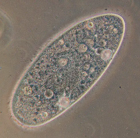
Paramecium
** Classification, Structure, Function and Characteristics
Paramecium is a unicellular organism with a shape resembling the sole of a shoe. It ranges from 50 to 300um in size which varies from species to species. It is mostly found in a freshwater environment.
It is a single-celled eukaryote belonging to kingdom Protista and is a well-known genus of ciliate protozoa.
As well, it belongs to the phylum Ciliophora. Its whole body is covered with small hair-like filaments called the cilia which helps in locomotion. There is also a deep oral groove containing not so clear oral cilia. The main function of this cilia is to help both in locomotion as well as dragging the food to its oral cavity.
Classification of Paramecium
Paramecium can be classified into the following phylum and sub-phylum based on their certain characteristics.
- Phylum Protozoa
- Sub-Phylum Ciliophora
- Class Ciliates
- Order Hymenostomatida
- Genus Paramecium
- Species Caudatum
Being a well-known ciliate protozoan, paramecium exhibits a high-level cellular differentiation containing several complex organelles performing a specific function to make its survival possible.
Besides a highly specialized structure, it also has a complex reproductive activity. Out of the 10 total species of Paramecium, the most common two are P.aurelia and P.caudatum.
Structure and Function
1. Shape and Size
P. cadatum is a microscopic, unicellular protozoan. Its size ranges from 170 to 290um or up to 300 to 350um. Surprisingly, paramecium is visible to the naked eye and has an elongated slipper like shape, that’s the reason it’s also referred to as a slipper animalcule.
The posterior end of the body is pointed, thick and cone-like while the anterior part is broad and blunt. The widest part of the body is below the middle. The body of a paramecium is asymmetrical. It has a well-defined ventral or oral surface and has a convex aboral or dorsal body surface.
2. Pellicle
Its whole body is covered with a flexible, thin and firm membrane called pellicles. These pellicles are elastic in nature which supports the cell membrane. It's made up of a gelatinous substance.
3. Cilia
Cilia refers to the multiple, small hair-like projections that cover the whole body. It is arranged in longitudinal rows with a uniform length throughout the body of the animal. This condition is called holotrichous. There are also a few longer cilia present at the posterior end of the body forming a caudal tuft of cilia, thus named caudatum.
The structure of cilia is the same as flagella, a sheath made of protoplast or plasma membrane with longitudinal nine fibrils in the form of a ring. The outer fibrils are much thicker than the inner ones with each cilium arising from a basal granule. Cilia have a diameter of 0.2um and helps in its locomotion.
4. Cytostome
It contains the following parts:
- Oral groove: There is a large oblique shallow depression on the ventrio-lateral side of the body called peristome or an oral grove. This oral groove gives an asymmetrical appearance to the animal. It further extends into a depression called a vestibule through a short conical funnel. This vestibule further extends into the cytostome through an oval-shaped opening, through a long opening called a cytopharynx and then the esophagus leads to the food vacuole.
- Cytopyge: Lying on the ventral surface, just behind the cytostome is the cytopyge also called a cytoproct. All the undigested food gets eliminated through the cytopyge.
- Cytoplasm: Cytoplasm is a jelly-like substance further differentiated into the ectoplasm. The ectoplasm is a narrow peripheral layer. It is a dense and clear layer with an inner mass of endoplasm or semifluid plasmasol that is granular in shape.
- Ectoplasm: Ectoplasm forms a thin, dense and clear outer layer containing cilia, trichocysts, and fibrillar structures. This ectoplasm is further bound to pellicle externally through a covering.
- Endoplasm: Endoplasm is one of the most detailed parts of the cytoplasm. It contains several different granules. It contains different inclusions and structures like vacuoles, mitochondria, nuclei, food vacuole, contractile vacuole etc.
- Trichocysts: Embedded in the cytoplasm are small spindle-like bodies called trichocysts. Trichocysts are filled with a dense refractive fluid containing swelled substances. There is a conical head on the spike at the outer end. Trichocysts are perpendicular to the ectoplasm.
5. Nucleus
The nucleus further consists of a macronucleus and a micronucleus.
- Macro Nucleus: Macronucleus is kidney like or ellipsoidal in shape. It's densely packed within the DNA (chromatin granules). The macronucleus controls all the vegetative functions of paramecium hence called the vegetative nucleus.
- Micro Nucleus: The micronucleus is found close to the macronucleus. It is a small and compact structure, spherical in shape. The fine chromatin threads and granules are uniformly distributed throughout the cell and control reproduction of the cell. The number in a cell varies from species to species. There is no nucleolus present in caudatum.
6. Vacuole
Paramecium consists of two types of vacuoles: contractile vacuole and food vacuole.
There are two contractile vacuoles present close to the dorsal side, one on each end of the body. They are filled with fluids and are present at fixed positions between the endoplasm and ectoplasm. They disappear periodically and hence are called temporary organs.
Each contractile vacuole is connected to at least five to twelve radical canals. These radical canals consist of a long ampulla, a terminal part and an injector canal which is short in size and opens directly into the contractile vacuole. These canals pour all the liquid collected from the whole body of paramecium into the contractile vacuole which makes the vacuole increase in size. This liquid is discharged to the outside through a permanent pore.
The contraction of both the contractile vacuoles is irregular. The posterior contractile vacuole is close to the cytopharynx and hence contract more quickly because of more water passing through. Some of the main functions of contractile vacuoles include osmoregulation, excretion, and respiration.
Food vacuole is non-contractile and is roughly spherical in shape. In the endoplasm, the size of food vacuole varies and digest food particles, enzymes alongside a small amount of fluid and bacteria. These food vacuoles are associated with the digestive granules that aid in food digestion.
Characteristics
1. Habit and Habitat
Paramecium has a worldwide distribution and is a free-living organism. It usually lives in the stagnant water of pools, lakes, ditches, ponds, freshwater and slow flowing water that is rich in decaying organic matter.
2. Movement and Feeding
Its outer body is covered by the tiny hair-like structures called cilia. These cilia are in constant motion and help it move with a speed that is four times its body’s length per second. Just as the organism moves forward, rotating around its own axis, this further helps it to push the food into the gullet. By reversing the motion of cilia, paramecium can move in the reverse direction as well.
Through a process known as phagocytosis, the food is pushed into the gullet through cilia which further goes into the food vacuoles.
The food is digested with the help of certain enzymes and hydrochloric acid. Once the digestion is completed the rest of the food content is quickly emptied into cytoproct also known as the pellicles.
The water absorbed from the surroundings through osmosis is continuously expelled from the body with the help of the contractile vacuoles present on either end of the cell. P. bursaria is one of the species which forms a symbiotic relationship with photosynthetic algae.
In this case, the paramecium provides a safe habitat for the algae to grow and live in its own cytoplasm, however, in return the paramecium might use this algae as a source of nutrition in case there is a scarcity of food in the surroundings.
Paramecium also feeds on other microorganisms like yeasts and bacteria. To gather the food it makes use of its cilia, making quick movements with cilia to draw the water along with its prey organisms inside the mouth opening through its oral groove.
The food further passes into the gullet through the mouth. Once there is enough food accumulated a vacuole is formed inside the cytoplasm, circulating through the cell with enzymes entering the vacuole through the cytoplasm to digest the food material.
Once the digestion is completed the vacuole starts to shrink and the digested nutrients enter into the cytoplasm. Once the vacuole reaches the anal pore with all of its digested nutrients it ruptures and expels all of its waste material into the environment.
3. Symbiosis
Symbiosis refers to the mutual relationship between two organisms to benefit from each other. Some species of paramecium including P. bursaria and P. chlorelligerum form a symbiotic relationship with green algae from which they not only take food and nutrients when needed but also some protection from certain predators like Didinium nasutum.
There has been a lot of endosymbioses reported between the green algae and paramecium with an example being that of the bacteria named Kappa particles giving paramecium the power to kill other paramecium strains which lack this bacteria.
4. Reproduction
Just like all the other ciliates, paramecium also consists of one or more diploid micronuclei and a polypoid macronucleus hence containing a dual nuclear apparatus.
The function of the micronucleus is to maintain the genetic stability and making sure that the desirable genes are passed to the next generation. It is also called the germline or generative nucleus.
The macronucleus plays a role in non-reproductive cell functions including the expression of genes needed for the everyday functioning of the cell.
Paramecium reproduces asexually through binary fission. The micronuclei during reproduction undergo mitosis while the macronuclei divide through amitosis. Each new cell, in the end, contains a copy of macronuclei and micronuclei after the cell undergoes a transverse division. Reproduction through binary fission may occur spontaneously.
It may also undergo autogamy (self-fertilization) under certain conditions. It may also follow a sexual reproduction process in which there is an exchange of genetic material because of mating between two paramecia who are compatible for mating through a temporary fusion.
There is a meiotic division of the micronuclei during the conjugation which results in haploid gametes and is further passed on from cell to cell. The old macronuclei are destroyed and formation of a diploid micronuclei takes place when gametes of two organisms fuse together.
Paramecium reproduces through conjugation and autogamy when conditions are not favorable and there is a scarcity of food.
5. Aging
There is a gradual loss of energy as a result of clonal aging during the mitotic cell division in the asexual fission phase of growth of paramecium.
P. tetraurelia is a well-studied species and it has been known that the cell expires right after 200 fissions if the cell relies only on the asexual line of cloning instead of conjugation and autogamy.
There is an increase in the DNA damage during clonal aging specifically the DNA damage in the macronucleus hence causing aging in P. tetraurelia. As per the DNA damage theory of aging the whole process of aging in single-celled protists is the same as that of the multicellular eukaryotes.
6. Genome
Strong evidence for the three whole-genome duplications has been provided after the genome of species P. tetraurelia has been sequenced. In some of the ciliates including Stylonychia and Paramecium UAA and UAG are designated as sense codons while UGA as a stop codon.
7. Learning
There have been some ambiguous results yielded, based on different experiments regarding whether or not paramecium exhibits the learning behavior.
There was a study published in 2006 which showed that P. causatum can be trained to differentiate between levels of brightness through a 6.5 volts electric current. For an organism with no nervous system, this type of finding is cited as a strong possible instance for epigenetic learning or cell memory.
Return to learning about Ciliates
Return from Paramecium to Unicellular Organisms Main Page
Return to Kingdom Protista Main Page
Find out how to advertise on MicroscopeMaster!
![Paramecium Diagram by Deuterostome [CC BY-SA 4.0 (https://creativecommons.org/licenses/by-sa/4.0)], from Wikimedia Commons Paramecium Diagram by Deuterostome [CC BY-SA 4.0 (https://creativecommons.org/licenses/by-sa/4.0)], from Wikimedia Commons](https://www.microscopemaster.com/images/512px-Paramecium_diagram.png)


