Ciliates Microscopy
** Habitats, Characteristics & Reproduction
What are Ciliates?
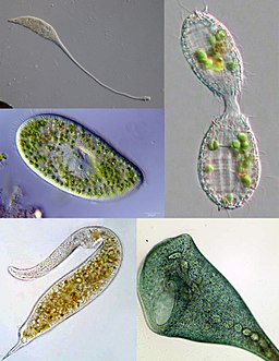
Essentially, ciliates are ciliated protozoans. As such, they are protists that belong to the super-group known as Alveolata along with dinoflagellates and apicomplexans. Because they are larger cells compared to other single-celled organisms, they feed on a number of other micro-organisms including bacteria and algae.
In addition to cilia (use for movement), ciliates also posses other short hair-like structures (membranelles) used for feeding.
* They are some of the most complex unicellular organisms.
Habitats
Ciliates are divided into free living and parasitic. Whereas free living ciliates (can live outside a host) can be found in just about any given environment, parasitic ciliates live in the body of the host.
Paramecium is an example of free living. Such paramecia as Paramecium caudatum can be found free living in fresh water bodies where they feed on bacteria.
Ciliates like Balantidium coli can be found in such host as human beings where they live as endoparasites and cause ciliary dysentery.
On the other hand, ciliates like Apospathidium terricola and Paraenchelys terricola can be found in soil. However, the concentration of these in soil is dependent on the amount of water in the soil. The higher the concentration of water in soil the more ciliates present.
* Their concentration will vary from one habitat to another depending on the conditions of the environment (nutrients, water, etc.)
Read more on Paramecium - Classification, Structure, Function and Characteristics
Microscopy
Ciliates like Paramecium can be viewed using the light microscope. To do this, a number of techniques can be used.
Requirements
- Sample of Paramecium species
- Microscope (bright field, phase contrast and dark field microscope)
- Microscope glass slide
- Microscope cover slip
- A dropper
- Vaseline
- Congo red dye
- Granular baker's yeast Bunsen burner
- Spring water
- Spatula
Wet Mount
There are a two ways through which the wet mount can be prepared for viewing under the microscope. To prepare Paramecium species for viewing, students may obtain the organism from pond water or culture the sample to increase their number.
Hanging Drop Technique
The hanging drop technique is the simplest method of preparing the sample for viewing. This simply involves suspending a drop of water on a cover slip. Here, the drop of water (pond water with the microorganism) is suspended on the underside of the cover slip, which is placed over a cavity of a glass slide.
Here, the water drop remains suspended between the cover slip and the glass slide (with a cavity) and viewed under the microscope at high power. When students use this technique, they will get an opportunity to view the microorganism moving fast across the field of view. Although it's transparent, students can identify them as they move about rapidly.
* Because paramecium are relatively large compared to other single-celled organisms, they can be easily identified using a brightfield microscope.
Wet Mount with Stained Yeast
The second technique involves preparing a wet mount of the sample with stained yeast. One of the main benefits of this technique over the former technique is that it causes the Paramecium to slow down, which makes it easier to view the organism and try identifying different structures.
This technique involves the following steps:
- Using a spatula, place a few grams of the baker's yeast in a beaker and add 100 ml of water (warm spring water) to hydrate the yeast
- Add about 0.3 mg/ml of Congo red dye and heat the suspension for about 10 minutes - This will reduce the suspension while concentrating the yeast.
- Using a dropper, place a drop of sample containing concentrated paramecium (concentration may be achieved by using a centrifuge) in contact with a drop of the stained yeast suspension.
- Put a little Vaseline on a cover slip and gently press the cover slip on the glass slide -The Vaseline allows for the retention of some air between the cover slip and the glass slide while also preventing the Paramecium from being crashed.
- Place the glass slide under bright field, dark-field and phase contrast microscope to compare how Paramecium cells appear.
* The dye used in the second technique (Congo red) is important because it serves as a pH indicator. As pH of the suspension changes from above 5 to below 3, color will change from red to blue.
* Compared to bright-field microscope, dark-field and phase-contrast microscopes will allow students to clearly identify the cilia at either ends of the cells as well as near the buccal cavity of the cells.
While the two techniques are important for viewing the cilia as well as a few other cell organelles of the organism, a bright-field microscope makes it easier to identify the food vacuole of Paramecia.
Characteristics
Cilia
As previously mentioned, ciliates are ciliated protozoa. This means that they are a form of protozoa with hair-like projections/organelles (cilia) originating from the cell cortex. These organelles are important for the organism given that they are used for movement.
According to studies, cilia are also used for crawling along surfaces as well as for attachment and sensation. Therefore, apart from helping the organism move from one region to another, they allow ciliates to sense any changes in their environments and therefore be able to respond effectively.
Compared to flagella present in other single-celled organisms, cilia are more numerous and short, and may cover the entire surface of the organism. Through their coordinated movement, they are able to rapidly move around more rapidly.
* While all have cilia, some use cilia to crawl (Aspidisca and Euplotes) while some are capable of swimming in water and are known as free-swimming ciliates (Paramecium etc).
Compared to other single-celled organisms, ciliates possess two nuclei; micronucleus and a larger macronucleus - The micronucleus consists of two copies of each chromosome making it a diploid nucleus.
Depending on the ciliate, there may be one or several micronuclei in a single cell. The macronucleus is larger than the micronucleus and contains short pieces of DNA (tens to thousands of copies). During cell division, the micronuclei often undergo mitosis while the macronucleus divided into two.
Oral Vacuole
Ciliates like Paramecia have a mouth-like structure refered to as an oral groove through which they feed. Modified cilia long the oral groove push the food particle through the cytopharynx (acting as the gullet) and into the food vacuole where the substrate is broken down. However, some lack an oral groove (mouth) and use absorption to feed/obtain nutrients.
Contractile Vacuole
Ciliates also have a contractile vacuole (Paramecia has an anterior contractile vacuole as well as a posterior contractile vacuole) that serves to collect and remove excess water from the cell.
When the concentration of water molecules is high inside the cell, they move into the contractile vacuole (which has higher ion concentration) and ultimately removed from the cell. This process allows the cell to maintain osmotic pressure and ionic balance while also preventing the cell from bursting due to excess water in the cell.
Reproduction
Ciliates may reproduce sexually (conjugation) or asexually (fission).
During conjugation (sexual reproduction), two ciliates come in contact with each other forming a cytoplasmic bridge between them. This is followed by a process known as meiosis of the micronuclei of either cell to produce haploid micronuclei.
Some of the haploid nuclei undergo disintegration while the remaining ones divide into two through a process known as mitosis in both cells.
One of either nuclei then moves to the other cell through the cytoplasmic bridge where it fuses with the micronuclei of the other cell to form a diploid nucleus ultimately forming a macronucleus once the cells separate. This is then followed by fission of the cell (while the macronucleus divided to two) to form two daughter cells. Each of the daughter cells will have a macronucleus and a micronucleus.
* During the fission phase of reproduction, the micronucleus of the cell go through mitosis (two diploid micronuclei) while the macronucleus divides into two. The cell then divides into two (splitting in to two daughter cells) with one of each macronucleus and micronucleus in each of the new cells.
Learn about Heterotrichs - Examples, Classification and Characteristics
Learn about Vorticella - Structure, Characteristics, Reproduction and Habitat
Learn about Tintinnids - The Species, Classification and Characteristics
Return to Cilia and Flagella page
Read more about Protozoa and Unicellular Organisms
Return from Ciliates Microcopy to MicroscopeMaster Research Home
References
George Karleskint, Richard Turner and James Small (2009) Introduction to Marine Biology.
https://biologywise.com/protozoa-classification-characteristics
Find out how to advertise on MicroscopeMaster!

![A labeled diagram of Paramecium By Deuterostome (Own work) [CC BY-SA 4.0 (https://creativecommons.org/licenses/by-sa/4.0)], via Wikimedia Commons A labeled diagram of Paramecium By Deuterostome (Own work) [CC BY-SA 4.0 (https://creativecommons.org/licenses/by-sa/4.0)], via Wikimedia Commons](https://www.microscopemaster.com/images/544px-Paramecium_diagram.png)


