What is the Difference Between Cytostome and Cytopharynx?
Overview
Both the cytostome and cytopharynx are specialized structures used for endocytosis.
They are commonly found in certain protozoans (e.g. Plasmodium falciparum) and vary in size and shape depending on the organism.
Together, they form the cytostome-cytopharynx complex through which food materials are transported into the cell and contained in vesicles.
Other examples of protozoans that possess the cytostome-cytopharynx complex include:
- Trypanosoma cruzi
- Trypanosoma brucei
- Balantidium coli
- Leishmania sp.
- Paratrypanosoma confusum
Cytostome
Also known as the micropore or cell mouth, the cytostome is located at the anterior end or near the anterior of the cell body. However, in some protozoans, it runs down the side of the cell body like a slit.
When viewed under electron micrographs, it looks like a dense ring around the neck of the cytopharynx (cytostomal invagination).
It varies in size between different organisms. For instance, in plasmodium falciparum, the inner ring of the structure (cytostome) measures about 90 nm in diameter. However, in Plasmodium species that infect reptiles and birds, the inner ring can be twice the size (170 to 200 nm in diameter).
In some literature, the cytostome is described as the opening on the cell membrane as well as the invagination. However, it's worth noting that the cytostomal structure only constitutes the opening region that leads to the invagination.
Using modified cilia, ciliates can move organic materials into the oral groove so that they can then be moved to the cytostome and eventually to the cytostomal invagination.
In some species, the region between the buccal cavity and cytostome is characterized by a sharp bend of the pellicle. A row of microtubular ribbons, also known as cytopharyngeal ribbons, originate from the bend and extend along the cytopharynx to the nascent food vacuole.
Also located at the cytostome (at the cytostomal lip) are box-like regions that consist of a single membrane covering the cytopharynx. The roof of these box-like regions is composed of microfibrils that are collectively known as the cytostomal cord. This cord rests over the end of the ribbons and runs along the entire length of the left edge of the cytostome.
Along the cytopharyngeal membrane, microtubular ribbons might be held in place by specialized microfilaments with arm-like structures.
Here, the microfilaments have been shown to link distal microtubules of individual ribbons to the membrane thus holding them in place. The bands, however, are not attached to the membrane at the end of the filaments. Rather, they curve away and extend into the cytoplasm.
In many species, another set of microtubules also arises at about the same level as the aforementioned band. Although they are located near the cytopharyngeal membrane and run parallel to the larger bands, they are not bound by filaments. However, they might be attached to the membrane by bridges.
The cytostome is also characterized by membrane-limited disks located along the length of the left lip. These uniquely shaped disks vary in size from 0.2 to 0.5 um in diameter. They also vary in thickness between cells. Whereas some of the disks appear flattened, about 40nm in thickness, others have been shown to be swollen or even spherical.
They are enclosed in a membrane similar to that of the plasma membrane and are often opaque. The opaque nature might be due to the leaflet-like layer at the lumen of the disk.
In the left lip of the cytostome, these disks are also associated with the cytopharyngeal microtubular band. Specifically, they aligned with the bands at a distance of about 40nm. The gap between them consists of finely filamentous material.
They are highly concentrated along the microtubular ribbons, near the cytopharyngeal membrane, and thus commonly found in the box-like structure beneath the cytostomal cord. Here, they are oriented in such a way that their edges are closest to the membrane. For some of the disks, however, their membrane is continuous with that of the cytopharyngeal membrane.
* The microfilaments extend about 1um along the band of microtubules.
* Food material received from the buccal cavity passes through the cytostome before reaching the cytopharynx. Therefore, the movement of food particles is one of the main functions of this structure. They have also been shown to play a role in the growth of membranes.
As mentioned, the disk-shaped vesicles are gradually oriented in a manner that ensures contact and fusion with the cytopharyngeal membrane. This is also suspected to increase the surface area for the development of food vacuoles.
Cytopharynx
The cytopharynx is also known as the cytostomal invagination (or cytopharyngeal basket).
It can be found in the majority of free-living protists and consists of the plasma membrane (invaginated plasma membrane). Unlike the cytostome, the cytopharynx has a tube-like morphology that originates from the posterior of the cytostome and extends into the cell body (towards the posterior end). However, like the cytostome, it varies in size between different species.
As compared to the cytostome, the cytopharynx is more elaborate. Generally, it is larger in size, has a more regular structure, and is also more compact.
Like the cytostome, the cytopharynx also has a circular cross-section.
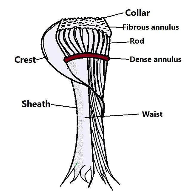
The lumen (inside the basket) is lined with a number of components that make up the cytopharyngeal wall. Aside from the rods surrounding the basket on the outside, the structure also consists of a collar ring at the top, the fibrous annulus below the collar, a dense annulus, a crest, and a sheath. The sheath is mostly uniform between the waist region and the annulus. However, it gradually declines towards the posterior end. The annulus, on the other hand, comprises broad and narrow segments.
Whereas the broader segments are located between the sides of the rods, the narrow ones pass around the outer surfaces of these rods. The crests, which run from the anterior region towards the waist region project outwards. However, the inner surfaces are usually attached to the sheath.
* The basket is also associated with three types of lamellae namely, the rod lamellae, the cytostomal lamellae, and the sub-cytostomal lamellae. Each of the lamellae mostly consists of a single, straight row of microtubules that is closely opposed by adjacent tubules.
Diagrammatic representation of a cytopharyngeal basket:
The Cytostome-Cytopharynx Complex
The cytostome and cytopharynx join to form the cytostome-cytopharynx complex.
As a product of the two structures, the complex consists of an opening at the surface of the plasma membrane as the cytostome as well as the invagination of the membrane which is the cytopharynx.
The general shape and structure of cytostome-cytopharynx change at different stages of cell development. For instance, early on in the G2 phase, the cytopharynx in T. cruzi has been shown to be shorter in length. However, as development continues during this phase, the complex appears fragmented with microtubules exhibiting a helical arrangement.
At the end of cytokinesis, the daughter cells possess a complete and functional cytostome-cytopharynx complex.
* Among members of the sub-genus Schizotrypanum, the cytostome-cytopharynx complex is only found in the epimastigote and amastigote forms.
Based on electron microscopy studies involving T. cruzi, the cytopharynx is supported by seven microtubules. Here, three of the tubules are located beneath the aperture membrane of the cytostome while the other four extend into the invagination from the flagella pocket membrane.
* The cytostome–cytopharynx complex in T. cruzi is between 6 and 11 um in length.
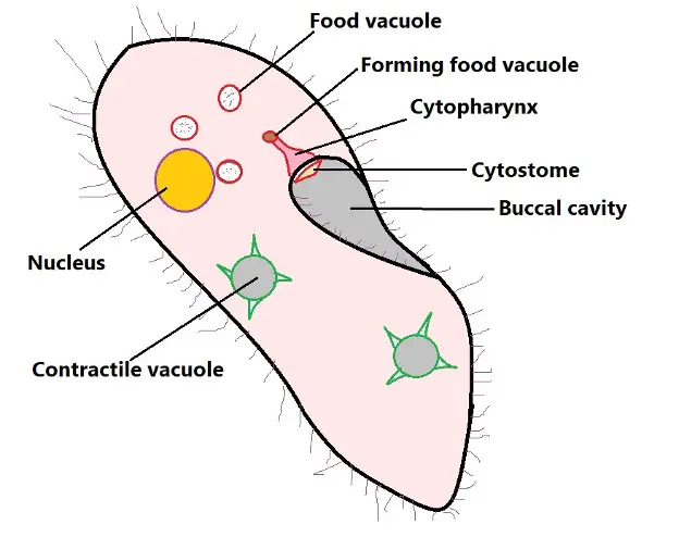
* As a unit, the cytostome–cytopharynx complex is located posterior to the buccal cavity. Whereas the cytostome leads food materials to the cytopharynx, the cytopharynx empties these materials into the budding food vacuoles at its posterior end.
Cytopharynx and Food Vacuoles
For the most part, food vacuoles are formed from the cytopharynx. As more food materials are received from the cytostome, the cytopharynx enlarges and a portion of it starts to constrict to form the food vacuole. Eventually, the newly formed vacuole buds off the base of the cytopharynx and moves to the posterior end of the cell.
At this stage (stage 1), the vacuoles do not contain acid phosphatase. These are lysosome enzymes responsible for the hydrolysis of organic phosphates. In paramecium, these vacuoles are bounded by acidosomes which are involved in the acidification process.
During this stage, the vacuole becomes increasingly acidic as the contents are condensed. Extra vacuolar membrane and excess fluids (fluids obtained along with food material) are also removed from the vesicle through a process known as pinocytosis and moved back to the cytopharynx.
In stage 2, the condensed food vacuole is surrounded by primary lysosomes which contain lysosomal enzymes. This causes the vacuole to enlarge and digestion continues. However, it becomes less acidic and might even test slightly positive for alkalinity. This continues in stage 3.
As digestion continues, the products are contained in smaller vesicles that bud off as secondary lysosomes and transported to the appropriate destination.
The vacuoles that enter stage 4 are much smaller in size and characterized by neutral ph. The lysosomal membrane is also retrieved before the vacuole enters the final stage.
In the final stage, the vacuole (containing waste products) fuses with the plasma membrane to excrete waste products. This occurs at a specific spot posterior to the buccal cavity known as the cytoproct.
Return to learning about the phylum Ciliophora
Return from "What is the Difference Between Cytostome and Cytopharynx?" to MicroscopeMaster Home
References
Alcantara, C. et al. (2017). The cytostome–cytopharynx complex of Trypanosoma cruzi epimastigotes disassembles during cell division. Published by The Company of Biologists Ltd | Journal of Cell Science (2017) 130, 164-176 doi:10.1242/jcs.187419.
Allen, R. D. (1974). Food Vacuole Membrane Growth with M Icrotubule-Associated Membrane Transport in Paramecium. Pacific Biomedical Research Center and Department of Microbiology, University of Hawaii.
Silva, N., Anna, C., Pereira, M., Souza, W. (2010). Endocytosis in Trypanosoma cruzi . 98 The Open Parasitology Journal, 2010, 4, 98-101
Tucker, J. B. (1968). Fine Structure and Function of the Cytopharyngeal Basket in the Ciliate Nassula. Department of Zoology, University of Cambridge.
Links
https://www.biologyonline.com/dictionary/cytopharynx
Find out how to advertise on MicroscopeMaster!


