What are Chromosomes?
** Relationship with DNA, Location and Structure
Definition: What are Chromosomes?
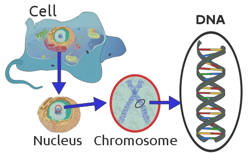
Discovered in the mid 19th Century, chromosomes are bundles of tightly packed DNA located in the nucleus (in eukaryotic cells). In prokaryotes, however, the chromosome exists as a circular DNA (located in the cytoplasm in the nucleoid) that can also be found in the plasmids. In eukaryotic cells, also, chromosomes can be found outside the nucleus in such organelles as the mitochondria.
In addition to carrying genetic material, chromosomes ensure that the DNA (which can extend to about 1.8 meters in length) is tightly compacted in a manner that allows it to fit inside the nucleus. Depending on the structure and the type of genetic material they contain, chromosomes can be divided into several categories making it easier to classify or group them.
Depending on the structure, there are four types of chromosomes that include:
- Metacentric
- Submetacentric
- Acrocentric
- Telocentric
A Brief History of Chromosomes
While Robert Hooke was among the very first scientists to identify the cell using a microscope in 1655 (Hooke also named these units "cells"), it was not until several years later that some more cell components would be discovered.
In 1719, for instance, Antonie van Leeuwenhoek identified a lumen at the central part of salmon red blood cells which were confirmed to be the nucleus by Robert Brown, a Scottish botanist, in 1831.
Although the nucleus had been identified by Franz Bauer, an Australian microscopist, in 1804, intensive studies of orchids (epidermis of orchids) under the microscope allowed Robert Brown to describe and name the organelle (He called this opaque area of the cell areola/nucleus)
These early studies laid the foundation for other scientists who were interested to learn more about the cell. In the mid-1800s, research studies by Matthias Schleiden and Matthias Schleiden, in 1839, and Rudolf Virchow, in 1858, led to the theory that the cell was the basic unit of life.
Later, in the 1870s, scientists like Oscar Hertwig realized that the nucleus was involved in inheritance through studies involving animal fertilization. However, it was in 1876 that Walter Fleming used staining techniques to stain and identify chromatin. This allows him to study cell division more closely in a process that he named mitosis.
In 1888, his colleague, Heinrich Wilhelm Gottfried von Waldeyer-Hartz continued studying the stained thread-like structures involved in cell division and named them chromosomes.
* Later studies allowed for the discovery and identification of the structure, composition as well as functions of chromosomes etc. For instance, in the early 1900s, an American biologist from the University of Columbia confirmed the presence of inherited factors in the chromosomes of Drosophila melanogaster in addition to discovering inherited diseases.
Composition and Structure of Chromosomes
Chromosomes are contained within the nucleus in eukaryotic cells, but can also be found in some of the other organelles such as mitochondria (where DNA is packed into a small circular chromosome).
In prokaryotes (e.g. bacteria), on the other hand, chromosomes form the nucleoid that is located in the cytoplasm given that these organisms do not have a membrane-bound nucleus.
Apart from the nucleoid (which is irregularly shaped), plasmids also contain chromosomes and are commonly referred to as extra-chromosomal DNA molecules because they are distinct from the chromosomal DNA of the organism.
Through microscopic techniques, the concentration of chromosomes (in the nucleus or the nucleoids) causes parts of the cell to have a dense, dark appearance which represents the regions of the cell that contains DNA material.
In organisms with several chromosomes (e.g. human beings have 23 pairs of chromosomes); the chromosomes vary in size and shape. For instance, in human beings, the first chromosome (chromosome number 1) has been shown to be the biggest in size and is about three times bigger than the 22nd chromosome.
As already mentioned, the chromosomes are made from the DNA molecule being tightly coiled and tightly packed. Here, the molecule of DNA is coiled around proteins known as histones that provide structural support. Therefore, molecules of DNA as well as histones are the main components of chromosomes.
DNA Molecule
Deoxyribonucleic acid (DNA) is the primary component of chromosomes and consists of two polynucleotide chains that coil around each other to form a double helix. It contains unique genetic codes and thus holds instructions that serve as the blueprint for proteins in the body.
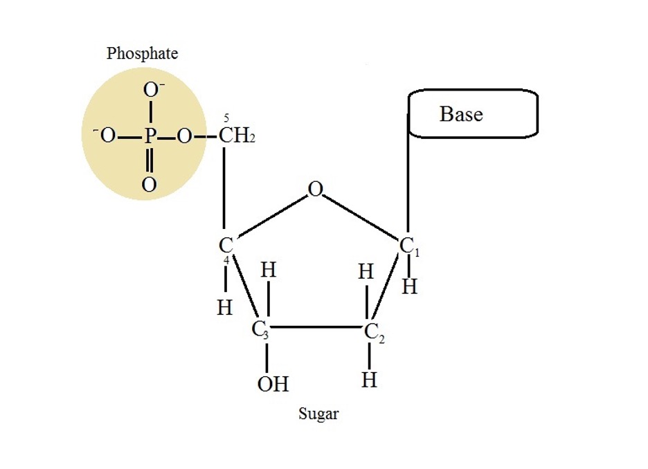
The DNA itself consists of compounds known as nucleotides. In the smallest chromosome, in human beings, a molecule of DNA is said to contain about 50 million pairs of nucleotides. For each strand of DNA, the nucleotides are made up of a nitrogenous base/nucleobase (adenine (A), guanine (G), cytosine (C), and thymine (T)), a sugar molecule as well as a phosphoric acid.
Nucleotides and DNA Molecule
As mentioned, a nucleotide consists of three main parts that include:
· A sugar - In DNA, the sugar group is composed of a 5-carbon sugar deoxyribose
· A phosphate - The phosphate is attached to the fifth (5th) carbon of the sugar
· The base - The base (nitrogenous base) is attached to the first carbon of the sugar. The bases are divided into two main categories that include the pyrimidines (C and T) and purines (A and G).
Purines and Pyrimidines
Both purines and pyrimidines are organic compounds that make up a DNA strand (polynucleotides). However, they have several differences that distinguish them from each other. Purines include guanine and adenine nucleobases.
Essentially, they are heterocyclic aromatic compounds that consist of a ring of pyrimidine fused to an imidazole ring. They are also characterized by two hydrogen-carbon rings as well as four atoms of nitrogen and have a melting point of 214 degrees C.
Pyrimidines, on the other hand, include thymine and cytosine (and uracil in RNA). They are described as heterocyclic aromatic compounds and consist of carbon and hydrogen. The structure of these bases is characterized by a single hydrogen-carbon ring as well as two atoms of nitrogen and has a melting point of between 20 and 22 degrees C.
* Whereas the catabolism of purines produce uric acid, the process results in the production of ammonia, beta-amino acids and carbon dioxide in pyrimidines.
* In a nucleotide, the phosphate group is joined to the pentose sugar through a phosphoester bond.
Nucleotides Polymerization
Nucleotides are the building blocks of a DNA strand. As such, they are the monomers that make polynucleotide chains to form the DNA. To form the polynucleotide chain, a bond is formed between the 3' carbon of the pentose of one nucleotide and the phosphate attached to the 5' (5th) carbon of a pentose of another nucleotide.
The bond formed between the two nucleotides is known as the phosphodiester bond/link given that it involves joining of the phosphate group to the adjacent carbon through an ester link. This occurs through a process known as condensation where a water molecule is produced.
Here, the hydroxyl group of a phosphate (in one of the nucleotides) reacts with the hydroxyl group attached to the carbon of the sugar of the other nucleotide (3rd carbon) to release a water molecule as the phosphodiester linkage is formed between the nucleotides. As the process is repeated with new nucleotides being added to the chain, a polynucleotide chain is formed and serves as the basis of a DNA strand.
* In this arrangement, the sugar-phosphate link acts as the backbone of the polynucleotide chain.
Base Pairing
In a chromosome, DNA is a double-stranded molecule (double helix). As such, it consists of two strands that wind around each other into what resembles a helical spring (like a twisted ladder). For the double-stranded molecule to be formed, each nucleotide in the DNA strand has to pair/link with another nucleotide (of the second DNA strand).
As mentioned, the phosphates and sugar make up the backbone of the structure. As such, they are located on the sides of the structure. This allows the nitrogenous bases, which are exposed, to join or pair-up with the bases of the second DNA strand thus forming a double-stranded molecule. With the phosphate and sugar forming the backbone of the structure, the bases bind and form the inner part of the double helix.
In the double helix, each of the base pair consists of two complementary nucleotides where a purine pairs with a pyrimidine. Here, the complementary relationship between the base pairs has been attributed to their shape which allows two particular bases to bind together through hydrogen bonds.
Here, Adenine and Thymine are bound by two hydrogen bonds while Cytosine and Guanine link through three hydrogen bonds. These bonds (hydrogen bonds) are weaker compared to other chemical bonds and thus allow the two strands to easily separate. This is particularly important given that DNA strands have to separate from time to time for transcription and replication to take place.
* The twisted geometry of a DNA helix is the result of base pairing. Here, a base at the 5' end of one of the strands is paired with a base located at the 3' of the second strand in an anti-parallel orientation. This arrangement causes the helix to twist and thus resemble a twisted ladder. This is also important in that it helps compact DNA.
* In some cases, a base or several bases can protrude from the double Helix. This is known as base flipping. Such bases can be removed by such enzymes as helicases.
DNA Packaging
DNA molecules are long and thus have to be packaged into chromosomes that can fit in the nucleus (in eukaryotes). Here, the strands of DNA are found around proteins known as histones to form nucleosomes that coil to form chromatins.
In some cases, however, the DNA is wound around non-histone chromosomal proteins. Based on molecular studies, researchers have shown that each of the nucleosomes consists of DNA strands wrapped around an average of eight (8) histones.
In addition to the eight histones, an additional histone is bound to the nucleosome as well as a linker DNA. As the nucleosomes and linker DNA continue to coil and form the chromatin fibers, DNA becomes increasingly compact.
Several steps are involved in the assembly of chromatins.
These include:
· Deposition of the (H3-H4)2 histone tetramer onto the DNA - This results in the formation of the sub-nucleosomal particle
· Maturation - The maturation step involves the use of ATP to form a nucleofilament through the spacing of nucleosome cores
· Folding of the nucleofilament following the incorporation of linker histones
· Folding and organization
* When they are formed, chromatins start folding in order to form chromosomes - A chromosome is therefore a complex of a double-stranded DNA and the packaging proteins.
Main Parts of a Chromosome
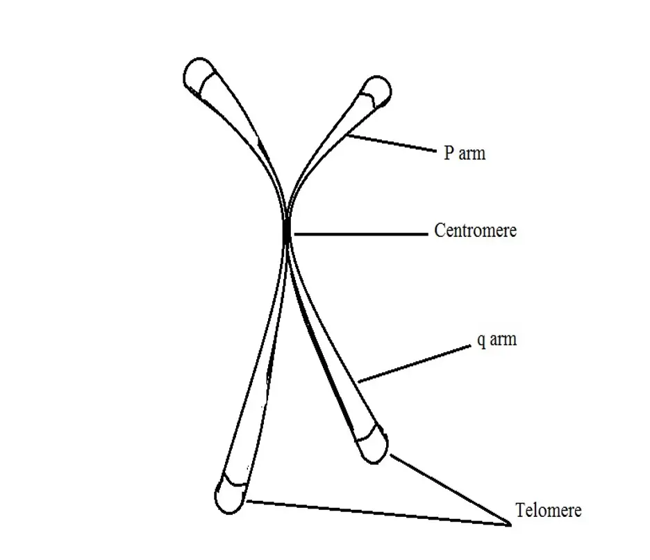
There are three main parts of a chromosome that include:
· Centromere - This is the constricted part of a chromosome that joins the chromatids together. This is also the point at which the microtubules attach to the kinetochore during mitosis (cell division).
· Telomere - Located at the end region of the chromatids, the telomere consists of repeated nucleotide sequences and serves to protect the chromosomes from deteriorating.
· Arm - A chromosome consists of two arms (p and q). These arms extend from the point of constriction (centromere) where the short arm is denoted p while the long arm is q. The arm of a chromosome gives the address of genes.
Based on the location of the centromere, chromosomes can be divided into four main categories.
These include:
· Metacentric chromosome - In a metacentric chromosome, the centrosome is centrally located. For this reason, the arms (p and q) appear to be of the same length.
· Submetacentric chromosome - A submetacentric chromosome is a chromosome in which the centromere is located near the center (but not at the center). For this reason, the p and q are of varying lengths.
· Acrocentric chromosome - In an acrocentric chromosome, the centromere is located near one end of the chromosome. As a result, some of the arms are significantly shorter than the others. Here, however, even the short arms are visible.
· Telocentric chromosome - In a telocentric chromosome, the centromere is located at one end of the chromosome. Unlike an acrocentric chromosome, only two arms are clearly visible in a telocentric chromosome.
Functions
Based on functions, chromosomes are also divided into two main categories.
These include:
Autosomes - In human beings, there are 23 pairs of chromosomes. 22 of these chromosomes are known as autosome chromosomes and are chromosomes other than the sex chromosomes. These chromosomes store a wide variety of genes that have unique functions in the cell.
Allosomes - Also known as sex chromosomes, allosomes (there is only one pair of allosome in the human genome) contain genes relating to the biological sex of an organism. Unlike autosomes, which are given numbers, allosomes are given letters (X and Y).
Learn about the Pentose Phosphate Pathway
Return to DNA under the Microscope
Return from Chromosomes to MicroscopeMaster home
References
Adrian T. Sumner. (2003). Chromosomes: Organization and Function.
Alberts B, Johnson A, and Lewis J, et al. (2002). Chromosomal DNA and Its Packaging in the Chromatin Fiber. Molecular Biology of the Cell. 4th edition.
Cooper GM. (2000). The Cell: A Molecular Approach. 2nd edition.
Rudi Appels, Bikram S. Gill, R. Morris, and C. E. May. (1998). Chromosome Biology.
Links
https://www.jove.com/science-education/10785/dna-packaging
https://www.news-medical.net/health/What-is-a-Chromosome.aspx
Find out how to advertise on MicroscopeMaster!


