Endocytosis
Definition, 3 Types, Active or Passive?, Vs Exocytosis
Definition
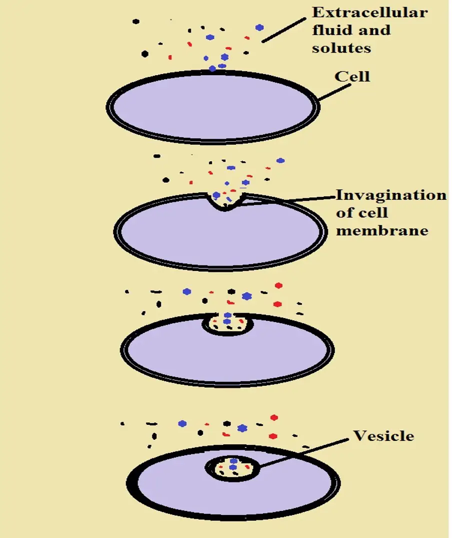
Endocytosis refers to the process through which materials or particles are internalized into the cell through the invagination of the cell membrane. Here, the cell membrane invaginates at the site where the particles to be internalized come in contact with the cell.
Invagination of the membrane results in the formation of a "pocket" around the particle/material which then buds off to form a vesicle inside the cell.
This type of transport is classified as active transport given that it serves to transport large particles/molecules (or a package of small particles/molecules) that cannot easily diffuse through the membrane. As a form of active transport, endocytosis also requires energy to move material/particles into the cell.
Endocytosis is an important process that serves a number of important functions including intake of nutrients required for normal cellular activities, engulfing invading microorganisms and other harmful substances, as well as engulfing old and damaged cells or parts of cells.
Active Vs Passive Transport
The movement of substances in and out of the cell without any energy input is known as passive transport. Generally, this involves the movement of molecules/ions from a region of high concentration (e.g. high concentration of molecules or ions in the extracellular environment) to an area of low molecule/ion concentration (e.g. inside the cell). The molecules or ions simply diffuse through the membrane to enter or leave the cell.
Active transport, on the other hand, requires the utilization of energy in the form of ATP for molecules/ions to be transported in or out of the cell. As mentioned, endocytosis is a type of active transport given that energy is required for molecules/substances to be transported into the cell.
ATP molecules have to bind to proteins on the cell which causes them to undergo a conformational change. This is an important activity that helps to cause the change in the shape of the membrane that contributes to the formation of vesicles. This allows for the molecules/substances of interest to be engulfed and transported into the cell.
There are three main types of endocytosis which include:
- Phagocytosis
- Pinocytosis
- Receptor-mediated endocytosis
Phagocytosis
Phagocytosis is one of the best-understood types of endocytosis. It's an important process used by a number of immune cells (macrophages, neutrophils, and dendritic cells) to engulf some invading microorganisms, dead or damaged cells as well as other particles that can cause harm to healthy cells.
Mechanism of Phagoctyosis
Before they can act (engulf given particles/materials etc), cells such as macrophages and neutrophils (phagocytes) first have to be activated. In the case of immune cells, such molecules as cytokines serve to activate the cell so that it can move to the site of infection to destroy the invading organism or damaged/dead cells.
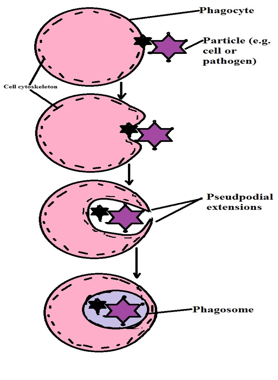
Given that they move in the direction of the activating molecule (e.g. cytokines), this type of movement is known as chemotaxis (moving in the direction of a chemical stimulus). Using pseudopodia (cytoplasmic extensions) on their surface, these cells can engulf and internalize the invading microorganism or damaged/dead cells at the infected site.
Generally, there are two ways through which phagocytes can identify the pathogen or infected/damaged cells. First, opsonization of the invading organism, given large molecules or infected cells by antibodies allow phagocytes to easily identify the pathogen or cell to be destroyed.
Here, opsonization can be thought of as tagging the pathogen or cell of interest so that it can be easily identified. As well, they can use receptors on their surface to identify various molecular structures known as pathogen-associated molecular patterns (PAMPs) located on the surface of pathogens, certain molecules, as well as infected cells.
For the most part, these structures are easily identified by phagocytes because of their characteristics. They may include nucleic acids, lipopolysaccharides, flagellin, and peptidoglycan layers, etc.
When these molecular structures come in contact with the receptors (pattern recognition receptors) located on the surface of phagocytes, they are recognized which activates a series of events that ultimately results in the molecule, bacteria, or cell, etc being engulfed and destroyed.
* An example of pattern recognition receptor is the Toll-like receptor 4 which recognizes lipopolysaccharide (LPS) on the surface of bacteria.
* Some of the receptors associated with phagocytes include mannose receptors (recognize microorganisms), scavenger receptors (recognize oxidized low-density lipoprotein as well as a variety of microbes), and receptors for opsonins (allow the cell to identify opsonized molecules).
Following the attachment of the molecule/cell/bacteria to the cell membrane of the phagocyte, a complex mechanism follows which results in its engulfment. First, receptor-initiated signals are integrated which in turn triggers membrane remodeling.
In general, this activation stimulates actin polymerization which in turn results in the formation of pseudopodial extensions.
Here, studies have shown that myosin has several important functions that include applying force to the actin filaments and plasma membrane among other several other components of the cell.
It's this action that influences the assembly of actin resulting in cytoskeletal changes and consequently the deformation of the plasma membrane as it forms pseudopodial extensions around the pathogen, molecule, or cell.
As the process continues, the particle is completely surrounded or engulfed in a phagocytic cup that buds off (detaches from the plasma membrane) to form a vacuole-like structure known as the phagosome. The phagosome then fuses with a lysosome (which contains several enzymes) to form a structure known as phagolysosome.
This is the site of particle killing and degradation and may involve several mechanisms that include:
· Oxidative bactericidal mechanism (accomplished by free radicals) - The particle is destroyed through oxidative damage
· Oxidative bactericidal mechanism (by lysosomal granules) - Lysosomal enzymes (may include lipases, hydrolases, etc) act on the particle to destroy it
· Non-oxidative bactericidal mechanism - Given that oxygen is not required for this process, the particle may be destroyed by granule enzymes or through the action of nitric oxide
* Phagocytosis is primarily characterized by actin polymerization which ultimately results in the deformation of the plasma membrane during the formation of pseudopodial extensions.
* Following the destruction and degradation of the particle (cell, molecules, or pathogen) in the phagolysosome, the contents are removed from the cell through exocytosis. This process is different from phagocytosis in that it involves the removal of substances from the cell to the extracellular environment.
Pinocytosis
Discovered in 1931 by Warren Lewis, pinocytosis is a type of endocytosis through which the cell takes in fluid (as well as dissolved solutes) from the extracellular environment into the cell. Pinocytosis is associated with a number of cells including liver cells, kidney cells, as well as epithelial cells.
Also known as cell drinking or cell sipping, pinocytosis allows these cells to take in various nutrients that may be present in the extracellular environment as well as cell signaling molecules, etc.
For this reason, pinocytosis and phagocytosis vary on the basis of function. However, differences can also be identified based on how both mechanisms work. Here, however, this mechanism also serves to transport small solutes in bulk across the cell membrane. As such, it also requires energy to accomplish this task.
Pinocytosis Process
While pinocytosis is a continuous process, like phagocytosis, it occurs in response to a molecule of interest. These molecules may include sugars, ions, proteins, or fats, etc in the extracellular matrix/extracellular fluid. In the extracellular fluid, these molecules first bind to cell receptors located on the cell membrane.
While receptors are involved in this process, it's different from phagocytosis and receptor-mediated endocytosis by the fact that these receptors (involved in pinocytosis) are not specific for a single molecule. For this reason, they can be activated by several types of molecules in the extracellular fluid to initiate the invagination process of the plasma membrane.
Following the binding of the molecules in the extracellular fluid to the receptors located on the cell membrane (of cells involved in pinocytosis), a signal is sent that triggers the invagination process.
Unlike phagocytosis which is dependent on actin polymerization, a protein known as clathrin is involved in the formation of vesicles in pinocytosis.
Here, the clathrin proteins are located in the cytoplasmic side of the cell membrane where they form a bristle-like structure. On this side of the cell, clathrins, which consists of clathrin adaptors, are associated with the receptor molecule. This allows the protein to receive the signal following binding of molecules in the extracellular matrix to the cell receptor.
While clathrin is involved in the formation of clathrin-coated vesicles, this process also requires such proteins as members of the Bin/Amphiphysin/Rvs superfamily (actin-binding proteins) among others to form the pit (during invagination of the plasma membrane).
Studies have shown the total areas of these pits to make up about 2 percent of the total area of the plasma membrane. Unlike phagocytes which engulf molecules by forming pseudpodial extensions, pinocytosis achieves this through the formation of clathrin-coated-pits.
The invagination of the plasma membrane surrounds the fluid and solutes with the vesicle finally pinching off from the cell membrane. The vesicle is made up of part of the cell membrane. This is particularly important in that the membrane protects the cell from contents of the vesicle which may be harmful to the cell.
As is the case with phagocytosis, the vesicle formed during pinocytosis fuses with lysosome which allows contents of the vesicle to be digested by enzymes.
* While this section has highlighted clathrin-mediated pinocytosis, this process may also be caveolin-dependent or even independent of the two proteins (caveolin and clathrin).
There are two types of pinocytosis which includes:
· Macropinocytosis - Characterized by smaller vesicles and is Caveolin-dependent
· Micropinocytosis - Characterized by relatively larger vesicles and involves the rearrangement of actin filaments
Phagocytosis VS Pinocytosis
While both phagocytosis and pinocytosis are types of endocytosis that function by engulfing material/molecules from the extracellular environment, the two have several important differences that make it possible to distinguish between them.
These include:
· During phagocytosis, pseudopodial extensions are formed around the molecule/cell/pathogen so that it can be internalized. This is different from pinocytosis where the plasma membrane invaginates to form vesicles allowing for fluids and solutes to be internalized.
· The material engulfed during phagocytosis are larger in size compared to those engulfed through pinocytosis.
· Phagocytosis is dependent on receptors that are highly specific while receptors involved in pinocytosis are not very specific.
· Pinocytosis is associated with different types of cells in the body and is a continuous process while phagocytosis is common with some immune cells among a few other cells and only occurs in response to given pathogens, molecules or when cells are damaged/dead.
· Phagocytosis allows cells to destroy given molecules while pinocytosis is mostly involved in the intake of signaling molecules and some nutrients.
Receptor-mediated Endocytosis
The third type of endocytosis is known as receptor-mediated endocytosis. This type of endocytosis serves to bring various materials needed by the cell. For instance, they may help in the importation of vitamins, hormones, and sugars among other macromolecules that a cell may need.
Unlike the other two types of endocytosis, receptor-mediated endocytosis is more specific given that receptors located on the cell surface only recognize and bind to specific molecules. Moreover, it has been shown to be more complex compared to the other types of endocytosis as it involves a number of important stages.
* Receptor-mediated endocytosis (RME) is also referred to as clathrin-dependent endocytosis.
Mechanism
In receptor-mediated endocytosis, the vesicles are formed at the site of the plasma membrane where the receptors are located. Here, binding of ligands to the receptors results in the formation of vesicles, a process known as stimulated endocytosis.
Recent studies have shown that in some cases, a ligand is not necessary for internalization of the receptors. In this case, internalization may occur in the presence or absence of ligands.
This type of internalization is known as constitutive endocytosis. This section, however, will focus on a scenario where a ligand binds to the receptors so that it can then be transported into the cell.
Following the binding of the ligand to the receptor, a signal causes a phospholipid component (PIP2- Phosphatidylinositol 4, 5-bisphosphate) to bind to the adaptor protein (AP2). As a result, the adaptor protein undergoes conformation changes.
This is important as the change in structure of the adaptor protein allows it to bind to the receptors on the cell membrane and recruit clathrin (the protein involved in the formation of coated vesicles).
Here, it's important to keep in mind that a clathrin molecule is a three-legged molecule consisting of three heavy chains and three light chains. Because of its structure, it can form complexes that take the shape of a sphere. For this reason, the clathrin molecules gradually form a spherical shape as they bind to more adaptor proteins on the cell membrane.
As a result, a spherical vesicle starts curving on the inside part of the cell as clathrin binds to more proteins. Ultimately, this results in the formation of a vesicle that is ready to be separated from the cell membrane. Once the vesicle has fully developed, dynamin, which is a GTPase acts as a pair of molecular scissors that separates the vesicle from the cell membrane.
At this point of receptor-mediated endocytosis, the vesicle consists of a number of molecules including the ligand, protein receptors, as well as adaptor proteins. For this reason, these components have to be sorted.
The first phase of sorting involves fusion of the vesicle with an early endosome. Because they contain proton pumps, the early endosomes tend to be slightly acidic. For this reason, binding of the vesicle to the endosome results in the ligands and receptors being separated.
Some of the receptors are then packaged into another endosome (recycling endosome) so that they can be recycled (transported to the plasma membrane for re-use). As well, the early endosome consisting of ligands and a few remaining receptors continues to mature as it develops into a late endosome.
Once the endosomes mature, they fuse with lysosome which contains various enzymes. Following fusion between the two, the enzymes degrade components of the endosome into simpler components. The products of enzyme degradation are then released into the cytoplasm where they can be used by the cell for various functions.
Following this degradation process, only the lysosome remains. Again, the lysosome fuses with a new late endosome, and the process is repeated.
* The maturation process of the endosome involves several important steps. For instance, parts of the endosome membrane that contain proteins to be degraded curve inwards thus allowing these proteins to be accessible to the degrading enzymes.
Secondly, a process known as glycosylation results in the formation of a protective layer around the endosome known as glycocalyx. This is an important layer that prevents the degradation of the endosome membrane thus preventing lysozymes from being released into the cytosol where they would otherwise degrade various components of the cell.
Lastly, the internal environment undergoes acidification (through the action of the proton pumps) which makes them highly acidic. This is important given that lysosomal enzymes normally function under acidic conditions.
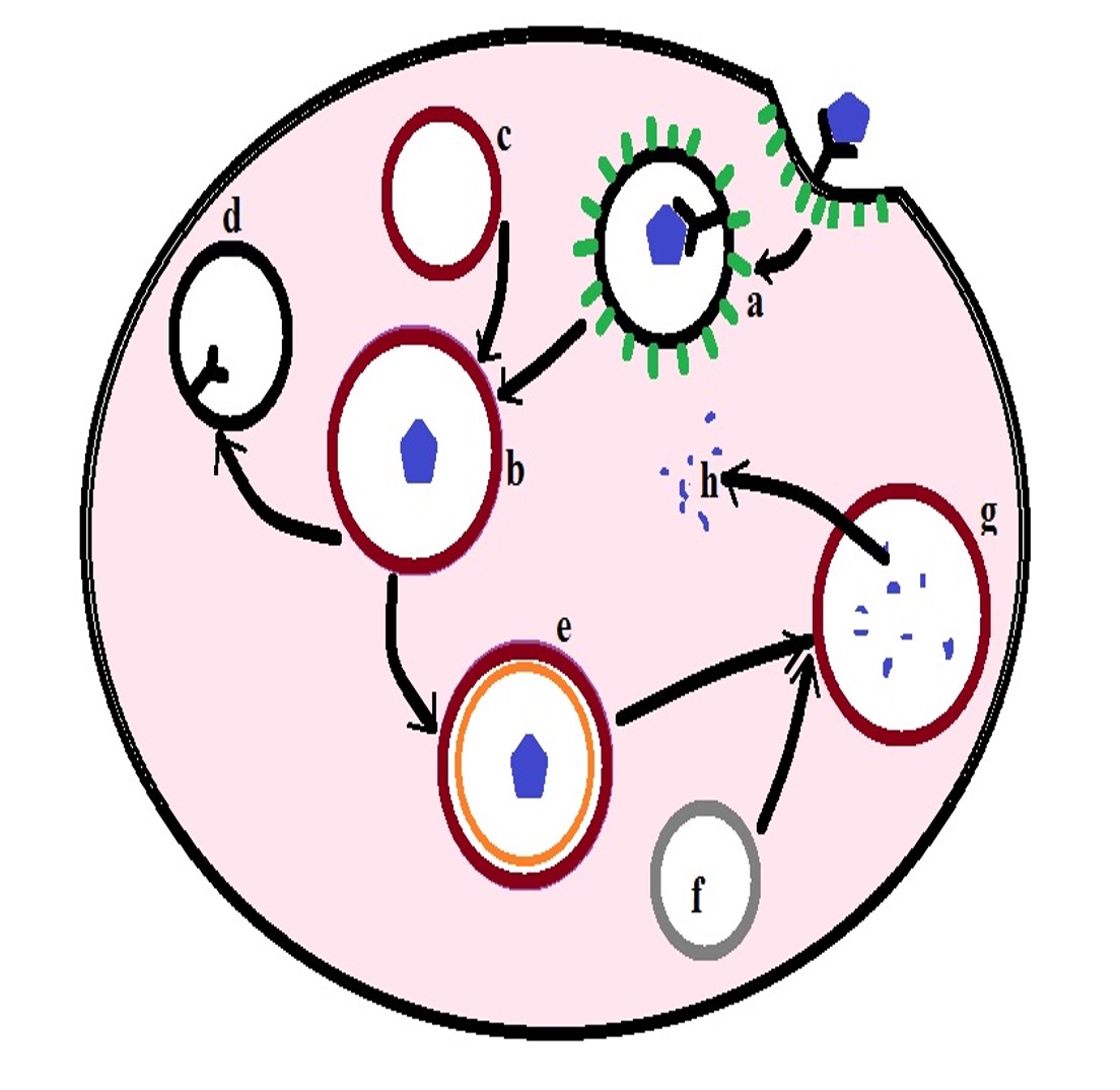
a- coated vesicle
b- early endosome
c- endosome (endosome fuses with coated vesicle to form the early endosome)
d- recycled receptors
e- late endosome
f- lysosome (lysosome will fuse with late endosome)
g- degradation of late endosome contents
h- degraded contents are released into the cytosol
Differences between Endocytosis and Exocytosis
The three types of endocytosis involve the movement of substances into the cell from the extracellular environments. This serves a number of important functions including the importation of nutrients and signaling molecules as well as the destruction of damaged and dead cells and invading pathogens among a few other functions. This is different from exocytosis which involves the transport of material out of the cell.
In exocytosis, substances/material to be removed from the cell are first packed into a vesicle by the Golgi body/Golgi apparatus. The vesicle then moves to and fuses with the cell membrane of the cell. This is easily achieved because the vesicle membrane has similar characteristics in structure to the cell membrane of the cell.
Following the fusion of the vesicle membrane and the plasma membrane, contents of the vesicle are released to the extracellular environment. In some cases, however, the vesicle may release its content and then return to the cell.
While exocytosis allows waste material to be packaged and removed from the cell, it also allows various molecules (e.g. protein) to be transported out of the cell so that they can be used elsewhere.
These molecules can be used to send signal molecules (e.g. hormones) to other cells which allows them to respond appropriately.
Therefore, one of the biggest differences between endocytosis and exocytosis is the fact that whereas endocytosis is the process of important molecules/pathogen entering into the cell, exocytosis serves to remove material out of the cell. However, they can also work together given that some of the material imported into the cell through endocytosis is modified before being removed from the cell through exocytosis.
Do animal cells have vesicles?
Return to Endocytosis Vs Exocytosis
Return to learning about Endosomes
Return from Endocytosis to MicroscopeMaster home
References
Alan L. Schwartz. (1995). Receptor Cell Biology: Receptor-Mediated
Endocytosis.
Cooper GM. (2000). Endocytosis: The Cell: A Molecular Approach. 2nd edition.
Eric J. Brown and Hattie D. Gresham. Cell Biology Of Phagocytosis.
Nicholas H. Battey, Nicola C. James, Andrew J. Greenland, and Colin Brownlee. (1999). Exocytosis and Endocytosis.
Links
https://www.jove.com/science-education/10708/receptor-mediated-endocytosis
https://www.sciencedirect.com/topics/immunology-and-microbiology/pinocytosis
Find out how to advertise on MicroscopeMaster!


