What are the Differences between Meiosis and Mitosis?
Introduction
One characteristic that is common among all living things is that they reproduce thus giving rise to a new generation of organisms. This allows them to pass down their genetic information to the successive generations. This is not only true for multicellular organisms but also for single-celled organisms.
For eukaryotic cells, mitosis and meiosis are two of the most common modes of cell division. Mitosis and meiosis have a number of similarities including sharing several steps during division, DNA duplication, and the fact that both processes produce more than one daughter cell.
While they share several characteristics, they also have a number of important differences related to their respective purposes.
Main Differences between Meiosis and Mitosis
Somatic vs. Germ Cells
One of the main differences between meiosis and mitosis is with regard to the types of cells involved in each process. Whereas meiosis occurs in germ cells, mitosis is the process of division in somatic cells. Essentially, somatic cells include cells that are not germline cells. As such, they include most cells that form the body of the organism.
Being the cells responsible for the formation of various body organs, somatic cells can be found in all parts of the body and are therefore not simply restricted to given regions. However, germline cells (gametes; sperm cells, and the ova) are confined to the reproductive organs.
While the gametes, like somatic cells, can undergo division, they are also capable of fusing in a process that produces a zygote during sexual reproduction. Somatic cells reproduce asexually (through mitosis) to produce two similar daughter cells. This not only allows for the replacement of lost or damaged cells but also for the growth of the organism.
Stages of Cell Division
Apart from the different types of cells involved in the two modes of cell division, one of the other important differences between meiosis and mitosis is with regard to the stages involved. While the stages involved are generally similar, the outcome is different.
This section will discuss the main stages involved in meiosis and mitosis
Mitosis
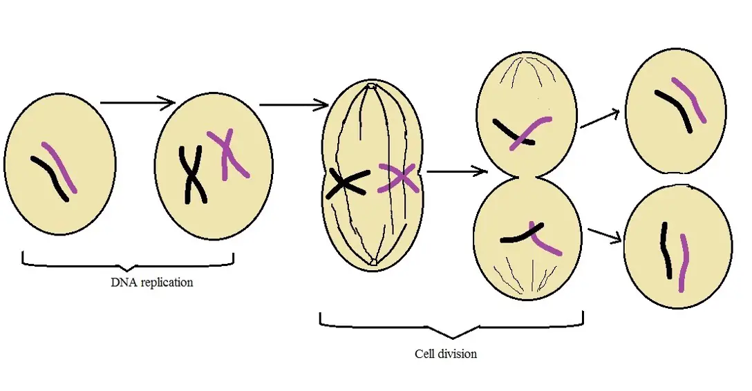
As mentioned, mitosis is the process through which somatic cells divide. This is an important cycle that results in the production of two similar daughter cells. Using human cells in culture, research studies have shown the typical cell to divide once every 24 hours (approximately).
Generally, mitosis is divided into two main phases that include:
Interphase - Interphase is the phase of the cell cycle in which the cell prepares for division. This phase is also divided into three main phases that include; The G1 (Gap 1) phase characterized by active metabolism and cell growth, the S (synthesis) phase characterized by DNA replication which results in doubling of the DNA, and the G2 (Gap 2) phase which is characterized by continued cell growth and synthesis of protein in preparation of the M (Mitosis) phase.
Here, it's worth noting that the G1 phase takes place following cell division. As such, it's an important stage where protein synthesis happens thus contributing to the cytosol content. During the S phase, proteins made during the G1 phase are used to produce histones on which DNA is wrapped around thus increasing the DNA content in the cell.
The number of chromosomes does not increase. In the G2 phase, organelles in the cell are duplicated which allows the cell to continue growing in preparation for mitosis. This is particularly important given that in addition to the DNA, cell organelles are also divided during the M phase.
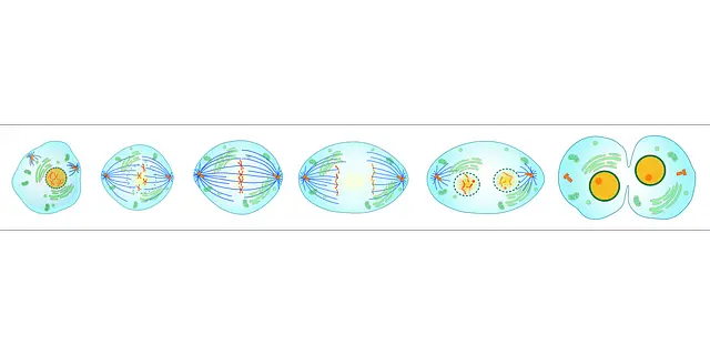
M Phase (Mitosis phase) - The M phase, or Mitosis phase, is the phase in which cell division takes place. While this phase of the cycle is divided into four main stages, the process of cell division is a progressive one. Some processes of one step are likely to continue in the next stage of cell division before they end for a new one to begin.
The main stages of the M phase includes:
Prophase - The prophase stage of M phase starts after the G2 phase. Although the cell continues to grow during the G2 phase, the DNA molecules are still intertwined. Prophase, on the other hand, is characterized by condensation of the DNA and proteins within the nucleus.
Here, the chromatins within the chromosomes coil becoming more compact which allows the chromosomes to become visible. This is because, during the condensation of the chromatic, the chromosomal material is untangled.
As a result, it's possible to identify the sister chromatids of the chromosome joined at the centromere. The prophase stage is also characterized by the formation of mitotic spindle (microtubules). They then start moving to the opposite poles of the cell.
By the end of this stage, it's possible to identify sister chromatids of the chromosomes as well as the mitotic spindles at the opposite poles of the cell. Some of the other cell components including the E.R, nuclear envelope, Golgi complexes and nucleolus, etc are not visible during Prophase.
Metaphase - Metaphase is the second stage of mitosis and is characterized by the total disintegration of the nuclear membrane. As a result, the chromosomes are released into the cytoplasm. Given that chromosome condensation is completed by the time the cell enters this stage, chromosomes are clearly visible which allows them to be studied with ease.
In addition to the sister chromatids, it's possible to identify centromeres as well as protein formation (disc-shaped) known as kinetochores around the centromeres.
The chromosomes released into the cytoplasm start to align at the central part of the cell. This is made possible by kinetochore microtubules that attach to the kinetochores and pull chromosome chromatids until they align at the equatorial plane (central part of the cell also known as the equator).
Here, alignment of the chromosomes at the equator is also known as the metaphase plate. This is an important process in that it's the checkpoint that ensures that the chromatids are ready to be separated.
Anaphase - Anaphase is the third stage of mitosis and is characterized by the separation of the sister chromatids that had aligned at the equator. Here, the chromatids are separated by the mitotic spindles which then start pulling them to the opposite poles of the cell.
Given that the sister chromatids are separated simultaneously, this process ensures that each of the daughter cells will eventually receive the same set of chromosomes (chromatids are the future chromosomes).
Telophase - Telophase is the fourth stage of mitosis and is characterized by the de-condensation of the new chromosomes. Here, a nuclear membrane also starts to form around the two sets of chromosomes thereby separating the DNA material from the cytoplasm. Because the chromosomes de-condense (uncoil), it becomes difficult to identify individual chromosomes.
Cytokinesis - While the other four stages of mitosis involve the division of DNA material, cytokinesis is the stage in which the cytoplasm of the cell starts to divide. As such, cytokinesis is generally viewed as the physical process of cell division. This is because of the fact that contractile rings formed around the equator start shrinking in a manner that causes the cell membrane to pinch, form a cleavage furrow, and ultimately separate the two daughter cells.
In plants, where cells have a cell wall, cell wall material is deposited at the center of the cell wall and continues growing until the daughter cells are separated.
* Although the stages of mitosis are subdivided into sub-stages (e.g. metaphase may be divided into prometaphase and metaphase), there are generally four stages of mitosis.
For the most part, this mode of cell division is limited to diploid cells and results in the production of similar daughter cells that contain the same genetic material as the parent. Daughter cells also become diploid cells.
* DNA replication occurs where the cell prepares for division. Following cell division (in the M phase), the daughter cell will contain the same number of chromosomes as the parent cell. These cells will then enter interphase where replication will increase DNA content in preparation for division.
Meiosis
Whereas mitosis occurs in somatic cells, meiosis is the mode of cell division in germline cells (gametes). These cells originate from specialized cells known as primordial germ cells. Like somatic cells, these cells are diploid and therefore have paired chromosomes. However, they give rise to daughter cells that are haploid through meiosis.
In organisms that reproduce sexually, meiosis is an important mode of cell division that produces gamete cells (eggs and sperm cells) that are ready to fuse during fertilization thereby restoring the diploid phase. As is the case with mitosis, meiotic cells first enter interphase before meiosis starts.
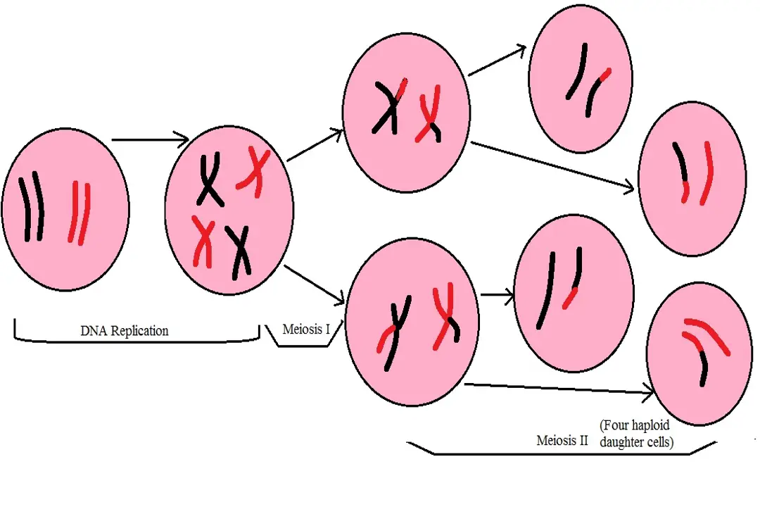
During interphase, the cell will grow followed by chromosome replication Chromosomes of the parent cell are replicated during the S phase producing identical sister chromatids. As such, interphase is the phase in which the cell prepares for meiosis. After the interphase phase, the cell enters two rounds of division which include meiosis I and meiosis II.
Meiosis I
Meiosis I is the first round of nuclear division. This phase is also known as the reduction division phase given that it ends up with half the number of the parent chromosomes.
As is the case with mitosis, meiosis 1 is divided into four main stages that include:
Prophase I - During the first round of division, prophase 1 is the first stage of division. As compared to the prophase stage in mitosis, this stage has been shown to be more complex.
It's also divided into several stages that include:
· Leptotene: This stage is characterized by compaction of the chromosomes which allows them to become visible.
· Zygotene: This phase of Prophase I is characterized by the pairing of the chromosomes. These chromosomes are commonly known as homologous chromosomes given that they contain identical genes and are generally the same size. Here, the two chromosomes pair tightly through a process known as synapsis. According to studies, this pairing has been shown to result in a complex that is known as a synaptonemal complex.
· Pachytene - This is the fourth stage of Prophase I and is characterized by clearly visible tetrads (paired chromosomes). This is also the stage in which genetic material is exchanged between the two chromosomes at sites known as recombination nodules. The process is mediated by an enzyme known as recombinase.
· Diplotene: This is the fourth stage of Prophase I and is characterized by separation of the homologous chromosomes. However, they do not completely separate at the crossover sites (chiasmata).
· Diakinesis: This is the last stage of Prophase I. In addition to the terminalization of the crossover sites, this stage is also characterized by condensed chromosomes, assembly of the meiotic spindles as well as breaking down of the nuclear envelope.
* While recombination also occurs during mitosis, it normally occurs between the sister chromatids during the replication phase. As such, it does not occur during the M phase (mitosis).
Metaphase I - During metaphase I, the bivalent chromosomes, that paired during Prophase I, start aligning at the equator. This is made possible by microtubules, located at the opposite poles of the cell, attaching to the homologous chromosomes at the kinetochore in a manner that helps line them up at the equator of the cell.
Anaphase I - Having attached to the homologous chromosomes, the microtubules contract which causes them to separate. While the spindle separates the homologous chromosomes, sister chromatids remain intact (joined together at their centromeres).
Telophase I - During telophase I, the nuclear membrane reappears around each set of chromosomes. In addition, the cytoplasm also divides ultimately resulting in the separation of the two daughter cells.
Each of these cells contains half the number of chromosomes of the original/parent cell. Given that recombination occurred during division, the daughter cells produced here are not genetically identical.
* This first round is also different from mitosis in somatic cells given that the number of chromosomes is half that of the parent cell.
Meiosis II
Before meiosis II starts, some cells enter a stage known as interkinesis. This is the stage in which some of the cells rest for a short period of time. However, this stage is also characterized by several activities such as further development of the nuclear membrane and disintegration of the spindle in some of the cells.
Unlike the first round of division, however, replication of the DNA does not precede Meiosis II. Meiosis II is also known as equational division.
As is the case with meiosis I, there are also four main stages in meiosis II, these include:
Prophase II - This stage is similar to the prophase stage in mitosis. Here, the chromosomes condense while the nuclear membrane breaks down. It's also characterized by the reformation of the spindle fibers in the event they disassemble during interkinesis.
Metaphase II - During metaphase II, chromosomes that were released into the cytoplasm gradually align at the central part of the cell (the equator) and are attached to the spindle fibers. This prepares for the separation of the sister chromatids.
Anaphase II - This is the stage in which spindle fibers (microtubules) attached to the sister chromatids at the kinetochore separates them.
Here, the chromatids are pulled to the opposite poles of the cell as the microtubules (spindle fibers contract). Given that each of the chromatids becomes a chromosome, cells that are ultimately produced will have the same number of chromosomes as the parent cells.
Telophase II - The chromosomes separated during anaphase are enclosed by a nuclear membrane/envelope. Ultimately, the cytoplasm also separates (cytokinesis) resulting in the production of four daughter cells.
* Given that two haploid cells enter meiosis II, meiosis II results in the production of four haploid daughter cells.
* Meiosis is particularly important because it results in the production of haploid daughter cells with half the genetic content of the original parent cell. This ensures that each of the gamete contains the proper number of chromosomes before fertilization occurs.
* In cases where meiosis does not divide chromosomes properly (particularly in meiosis I), then extra chromosomes may result in such genetic disorders as trisomy following fertilization.
* During meiosis, the DNA is replicated in preparation for meiosis I and II. At the end of meiosis II, the four daughter cells will have half the number of chromosomes that of the parent cells.
See also:
Return to Basic Components of Cell Theory
Return to learning about Cancer Cells
Return from "What are the Differences between Meiosis and Mitosis? to MicroscopeMaster home
References
Andreas Hochwagen. (2008). Meiosis.
Elizabeth R. Cregan. (2007). All About Mitosis and Meiosis.
Lynette Clark. (2016). Cell Cycle and Cell Division.
Links
https://www.technologynetworks.com/cell-science/articles/mitosis-vs-meiosis-312017
https://www2.le.ac.uk/projects/vgec/highereducation/topics/cellcycle-mitosis-meiosis
Find out how to advertise on MicroscopeMaster!


