Onion Root Tip Mitosis
Stages, Experiment and Results
Overview
Mitosis refers to a type of cell division (cell cycle) through which the cell (parent cell) produces two identical daughter cells. Unlike meiosis, which is also a type of cell division, mitosis results in the production of two diploid daughter cells. The two daughter cells contain the same number of chromosomes as the parent cell.
Given that the process results in the proliferation of cells, it's important for general growth and replacement of damaged cells (e.g. the wound healing process).
In general, mitosis occurs through several stages that include:
- Prophase (divided into prophase and prometaphase)
- Metaphase
- Anaphase
- Telophase
Because of the rapid rate at which onion root tips grow as a result of rapid cell division, it's possible to observe and identify the different stages of mitosis.
Experiment
For this section, the experiment will be divided into two main parts.
These include:
- Growing onion root tips
- Sample preparation
Growing onion roots
* Growing fresh root tips is recommended given that older or dried roots may produce poor results.
Requirements
- Uncut onions
- Clear glass or plastic jars
- Clean water
Procedure
- Pour clean water into the clean (and clear) glass/plastic jars - about three-quarters full.
- Carefully place the onion bulbs in the glass/plastic jars so that only the lower surface of the onion comes in contact with the water.
- In a case where the onions are too small, they can be supported using toothpick/splints.
- Let the onions rest on water for 3-4 days - only the lower surface (base from which roots emerge) should be in contact with water.
The following are diagrammatic representations of this setup:

Results
After 3 to 4 days, the onion roots will have grown to a length of about 1 inch.

Sample preparation
Requirements
· 70 percent alcohol
· Aceto-alcohol
· Blade
· Forceps
· Water
· Glycerin
· Stain - Aceto-carmine or Aceto-orcein
· Hydrochloric acid solution (1N HCL)
· Onion with freshly grown roots
· Stop clock
· Water bath
· Dissecting needles
Procedure
* Having a pair of gloves, safety goggles, and safety mask is recommended given that some of the chemical used are hazardous.
· Using a blade or scalpel, carefully cut one or several roots from the onion and place them in a petri dish (or any other small clean container)
· Prepare a water bath (about 55 degrees C) - The temperature should be maintained at 55 degrees C. This may be achieved by using a thermostatically controlled water bath
· Carefully pour the hydrochloric acid solution into a small bottle and place it in the water bath for about 15 minutes in order to warm the acid
· Using a pair of forceps, carefully pick one or two of the roots and place them into the bottle of warm hydrochloric acid for about 5 minutes - This serves to break down pectin and calcium pectate as well as other tissues in order to release individual cells
· Remove the roots from the bottle of acid using the forceps and rinse them in tap water several times and then place them in a clean petri dish
· Using a clean blade, sharp scissors or scalpel, cut off the tips of the roots (about 5mm in length)
· Using the pair of forceps, pick the root tips, and put them into a vial of stain (acetic orcein or aceto-carmine) - Ensure that the root tips are completely immersed in the stain
· Place a cap/lid onto the vial (ensure that the cap/lid has a pinprick hole) and place the vial in the water bath (at 55 degrees C) for about 5 minutes - This enhances the staining process
· Using the forceps, remove the root tips from the vial of stain and place them onto a clean microscope glass slide
· Using a dissecting needle, you can gently mash/squash the root tips to spread out the cells on the glass slide - This prevents several layers of cells from overlapping which would otherwise affect the quality of results
· Cover the sample (root tip) with a coverslip and gently press the coverslip down, then examine the slide under the microscope starting with low magnification
* For this experiment, a properly prepared slide should appear light pink due to the stain to almost colorless.
* Unused roots can be stored in 70 percent alcohol.
Results
An onion has a total of 8 pairs of chromosomes. This is especially beneficial for this experiment given that fewer chromosomes are slightly easier to see when they condense.
As mentioned, onion root tip cells divide rapidly as the roots elongate to absorb water and various minerals from the soil.
For this reason, it's possible to identify different stages of cell division by mitosis based on chromosomal distribution.
When viewed under the microscope, a properly prepared slide will yield the following results:
Under 10X magnification
Under 10X magnification, students will be able to observe several single layers of cells. Depending on how well the slide was prepared, the cells are spread out without any overlapping or with very little overlapping.
* Generally, a row of cells (single layer) may consist of between 2 and 5 cells.
The following is a diagrammatic representation of onion root tip cells under 10X magnification:

Higher magnification (stages of mitosis)
Under high magnification (100X-500X), it becomes possible to identify cells at different stages of mitosis.
These include:
Interphase
While this is not necessarily one of the main stages of mitosis, cells in this state can easily be identified by their prominent nucleoli.
Diagrammatic representation of an onion root tip cell during interphase:

During interphase, also known as the resting stage, the chromatin is not tightly packed. This allows for DNA to be copied (replication) as the cell prepares for cell division.
Generally, interphase is divided into three main phases that include:
· G1 (First gap phase) - Characterized by cell growth and normal cellular activities
· S phase (Synthesis) - This is the phase in which DNA is replicated so that the DNA content is doubled
· G2 phase (second gap phase) - During this phase, the cell prepares for division
Prophase
During this stage of mitosis, the nucleolus is still visible. However, the nucleus appears grainy due to chromosomal condensation.
The following is a diagrammatic representation of an onion root tip cell during prophase:

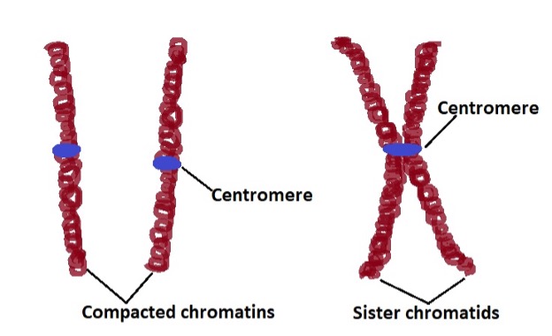
As mentioned, the DNA strands are uncoiled during interphase for transcription to occur. In this state, the DNA is said to be uncondensed. Here, this genetic material is also referred to as chromatin. During prophase, condensed DNA and some proteins form the chromatids which join to become chromosomes (X-shaped).
Sister chromatids, which contain the same genetic information, are attached at a region known as the centromere which gives the structure an X shape.The kinetochore, which is the site at which microtubules join the chromosomes is also located at the centromere.
* As the chromatins coil, it becomes increasingly compact which allows the chromosomes to become more visible when viewed under high magnification.
* It's also during prophase that spindle microtubules (mitotic spindle) start forming near the nucleus.
Prometaphase
Prometaphase is the second stage of mitosis (it has also been referred to as late prophase). This stage is characterized by increased condensation of chromosomes as well as the breakdown of the nuclear envelope (nuclear membrane).
The nuclear envelope, which consists of an inner and outer membrane, is stabilized by polymerization of lamin proteins (the nuclear lamina). Polymerization of the lamins and the consequent breakdown of filaments into lamin dimers results in the disassembly of the nuclear membrane.
Apart from increased condensation of the chromosomes and breakdown of the nuclear envelope, this stage is also characterized by the development of kinetochore around the centromere. Moreover, spindle fibers (kinetochore microtubules) have also been shown to start developing during prometaphase and permeate through the disappearing membrane to attach to the chromosomes at the kinetochore.
* This stage of mitosis is important because chromosomes (sister chromatids) have to be released from the nuclear membrane in order to be separated in the next stage.
Metaphase
In this stage of mitosis, chromosomes align along the equatorial plane of the cell (cell equator) so that the sister chromatids can be separated. During metaphase, chromosomes become more visible because of increased condensation as well as the fact that the nuclear envelope has disappeared.
Diagrammatic representation of a cell during metaphase:

Given that the nuclear membrane completely disappears during metaphase, the chromosomes appear in the cytoplasm. As well, the fully developed spindle fibers originating from the centrioles, located on opposite poles of the cell, attach to each of the sister chromatids which contribute to their alignment at the cell equator.
Based on microscopic studies, spindle fibers (microtubules made of proteins) have been shown to be about 25nm in diameter. While they originate from the centrioles and extend to attach to the sister chromatids, they have also been shown to be constantly forming because they are continually broken down.
As new components (building blocks) are added on one end of the microtubules and removed from the other, it has been suggested that this causes the microtubules to pull the centrioles which in turn contributes to the alignment of the chromosomes.

* In late metaphase, the pulling actions of the microtubules, as well as the centrioles, result in the kinetochores (the region at which spindle fibers attach to the chromosomes) facing different directions.
Anaphase
During anaphase, the chromosomes start separating and moving from the equatorial plate of the cell. At the end of this stage (late anaphase), the sister chromatids completely separate and reach the opposite poles of the cell.
In this stage, it's also possible to clearly identify the chromatids given that the process occurs in the cytoplasm.
Diagrammatic representation of a cell during late anaphase:

Based on microscopic studies, sister chromatids have been shown to separate and move to the opposite poles of the cell at a rate of between 0.2 and 4 um per minute. Initially, polymerization and depolymerization of microtubules was thought to result in the separation and movement of chromatids to the opposite poles of the cell.
As a result of the depolymerization process, microtubules shorten thus pulling apart the sister chromatids. As well, polymerization of some of the microtubules, those that extend from one pole of the cell to the other without attaching to the chromatids, causes them to grow in length thus pushing poles of the cell apart.
Based on recent studies, however, it has become evident that separation of sister chromatids during anaphase is the result of the actions of enzyme seperase as well as shortening of microtubules.
Here, the enzyme seperase has been shown to break down cohesin (a component of centromere that links sister chromatids).
Following the separation of sister chromatids, through the breakdown of the protein cohesin, shortening of microtubules (kinetochore microtubules/spindle) pulls the chromatids to the opposite poles of the cell.
* Some of the other factors suggested to contribute to the separation of sister chromatids include the pulling actions of astral microtubules (pulling the poles apart) while interpolar microtubules slide past each other.

Telophase
Telophase is the fifth stage of mitosis characterized by several key events. These include arrival of the chromosomes at the opposite poles of the cell, gradual breakdown of the spindle fibers as well as development of nuclear envelopes around each set of chromosomes (at the opposite ends/poles of the cells).
Diagrammatic representation of a cell during telophase (late telophase):
, Onion root tip mitosis. Credit: MicroscopeMaster.com")
%2C%20Onion%20root%20tip%20mitosis.%20Credit%3A%20MicroscopeMaster.com)
As nuclear envelopes develop around each set of chromosomes located at the opposite poles of the cell, two nuclei are formed in the cell. The DNA also starts to uncondense so that genetic material can be copied later.
Given that the spindle fibers are no longer required, they start to disassemble (break down) in early telophase and continue to do so in late telophase.
Cytokinesis
Cytokinesis refers to the process through which the cytoplasm separates as the cell divides into two identical daughter cells. Unlike animal cells, plant cells have a rigid cell wall that prevents the cell from easily pinching apart to form two identical daughter cells.
For this reason, the process of cytokinesis is different in these cells (plant cells). During late telophase and early cytokinesis, carbohydrate filled vesicles are released by the Golgi bodies and occupy the equator region of the cell. The vesicles continue fusing to form the cell plate which divided the cell into two.
Here, the carbohydrates contained in the vesicles form the middle lamella between the membrane of the two cells. The cellulose produced by the two new cells occupies the region between the middle lamella and cell membrane to form the primary cell wall for the two daughter cells.
Difference between Meiosis and Mitosis
Return to Onion Cells under the Microscope
Return from Onion Root Tip Mitosis to Microscopemaster home
References
Cooper GM. (2000). The Cell: A Molecular Approach. 2nd edition.
Donald, B. and Richard, J. (2018). Cell Division. An Atlas of Comparative Vertebrate Histology.
Lian, Y. and Chirop, M. (2016). Functional Cell Biology. Encyclopedia of Cell Biology.
Lodish H, Berk A, and Zipursky SL, et al. (2000). Microtubule Dynamics and Motor Proteins during Mitosis: Molecular Cell Biology. 4th edition.
Links
https://employees.csbsju.edu/ssaupe/biol115/cell_division.htm
https://www.yourgenome.org/facts/what-is-mitosis
Find out how to advertise on MicroscopeMaster!


MicroscopeMaster.com is a participant in the Amazon Services LLC Associates Program, an affiliate advertising program designed to provide a means to earn fees by linking to Amazon.com and affiliated sites.
Recent Articles
-
Deltaproteobacteria - Examples and Characteristics
Nov 01, 22 04:44 PM
 Deltaproteobacteria is a large group (Class) of Gram-negative bacteria within the Phylum Proteobacteria. It consists of ecologically and metabolically diverse members. Read more here.
 -
Chemoorganotrophs - Definition, and Examples
Oct 26, 22 05:01 PM
 Chemoorganotrophs also known as organotrophs, include organisms that obtain their energy from organic chemicals like glucose. This process is known as chemoorganotrophy. Read more here.
 -
Betaproteobacteria – Examples, Characteristics and Function
Oct 25, 22 03:44 PM
 Betaproteobacteria is a heterogeneous group in the phylum Proteobacteria whose members can be found in a range of habitats from wastewater and hot springs to the Antarctic. Read more here.