Pili and Fimbriae
Types, Function and Differences
Overview: What are they?
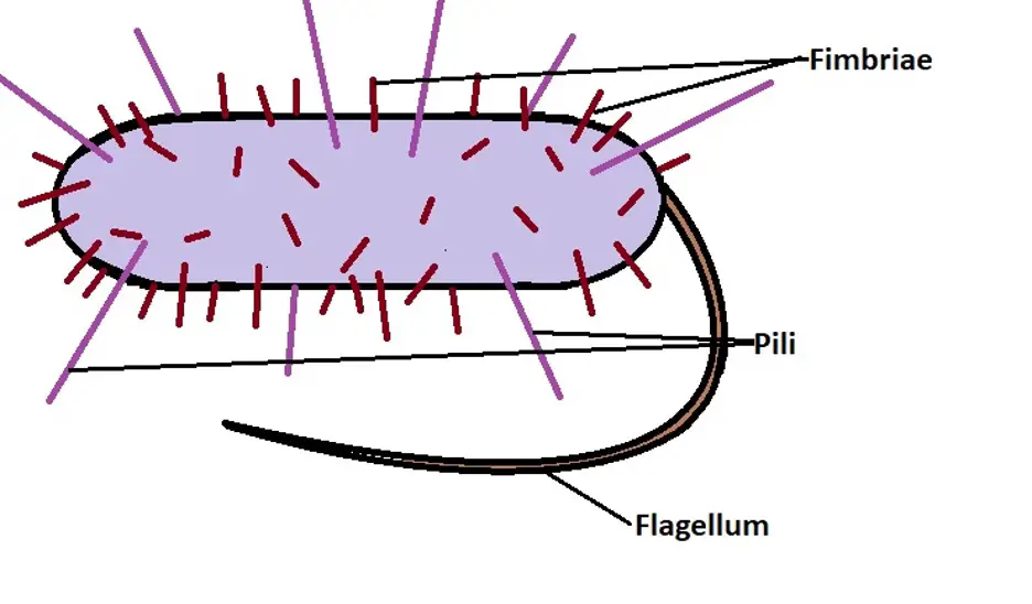
Pili and fimbriae are proteinaceous, hair-like structures/appendages that extend from the cytoplasmic membrane of a variety of bacteria. Compared to flagella, they are both shorter and thinner in size. However, they are also different from each other and have several functions.
While they can be found in some Gram-positive bacteria, they are some of the most common features of all Gram-negative bacteria.
See more on Gram positive and Gram negative bacteria.
Types of Pili
Pili (singular: Pilus) are longer in length and thicker when compared to fimbriae. They can be found in some Gram-positive species of bacteria and all Gram-negative bacteria as well as archaea.
Generally, there are two main types of pili.
These include:
Conjugative Pili
Also known referred to as sex pili in some books, conjugative pili are some of the most common bacterial pili. As the name suggests, these structures are involved in the transfer of genetic material from one cell to another.
Compared to the other pili, the F pilus (of F sex pilus) has been given more attention and is, therefore, better understood. Encoded by the F plasmid, the F pilus is found in "male" Gram-negative bacteria (F+).
Based on microscopic studies (using powerful electron microscopes), these appendages have been shown to be dynamic and therefore elongate and retract continuously.
With regards to structure, the pilus is a polymer that consists of the protein pilin (VirB2) and is also structurally similar to the F-like pilus pED208 - They both measure an average of 87 Å in diameter with an internal lumen that is about 28 Å in diameter.
Depending on the type of pili, the building blocks (proteins) may be arranged in a helical manner (five-start helical filaments) or in pentamer layers on top of each other.
Given that bacterial pili originate from the membrane, they also consist of phospholipids, and proteins, which form a protein-phospholipid complex. However, the lipid in pilus has been shown to be slightly different from the lipids in the membrane which is attributed to the binding of TraA to part of the phospholipid during pili formation.
* F-pili measure about 20um in length.
Functions of Conjugative Pili
As mentioned, conjugative pili are primarily involved in the transfer of DNA from one bacterial cell (male F+ ) to another (F-). This is why they are also referred to as sex pili.
While the process is not properly understood, it's suggested that single strands of DNA can pass through the hollow lumen of the pilus to be transported to the recipient cell.
Although this process was not widely accepted, some studies have shown that genetic material can be transferred over a distance (where the donor and recipient cells are not directly in contact with each other) through the F-pili.
* The F-pilus is also thought to play an important role in the identification of the recipient as well as promoting contact between the two cells, the donor and recipient cell, as it retracts.
Type VI Pili
Type IV pili are a type of Pili found in some Gram-positive bacteria (e.g., clostridia) and the majority of Gram-negative bacteria. They have several important functions ranging from their role in locomotion to DNA exchange.
Like conjugative pilus, type IV pili are tube-like structures that originate from the membrane. While they are strong structures, they are also flexible which allows them to effectively perform their functions.
Structurally, these pili (type IV pili) are also polymers that consist of pilin protein. Here, numerous copies of pilin (thousands of copies) are polymerized from subunits in the membrane to construct the filament (type IV pilus).
Although the polymerization of these pilin copies is not properly understood, it's generally believed to involve the assembly of ATPase as well as the core protein of the inner membrane.
* Pilins involved in the formation of type IV pili are divided into two main categories that include; type IVa pilins (characterized by 5 to 7 amino acid signal peptides and phenylalanine) and type IVb pilins which consist of relatively longer peptides and a hydrophobic residue.
As the pilus is constructed, a protein known as secretin is involved in the formation of oligomeric gated channel in the outer membrane of bacterial cells so that the pilus can pass through.
Here, PilN and PilO, which are pilus alignment subcomplex proteins associated with the inner membrane, also come in contact with secretin and channel the protein PilP which results in the formation of the periplasmic conduit so that the pilus can grow through.
These activities allow the pilus originating from the membrane to extend outwards into the outer environment.
* Type IV polymers also have a helical orientation with a diameter of about 6nm and 1um in length.
* Depending on the species, the pilin may be glycosylated or phosphorylated.
Functions of Type IV Pili
Compared to conjugative pilus, type IV pili have a number of functions that include:
Adhesion - Adherence is one of the primary functions of Type IV pili. In addition to attaching a bacterial cell to different types of surfaces, the filament also allows the cell to adhere to other bacteria.
The ability of bacteria to adhere to a variety of surfaces is made possible by the highly diverse sequence of pilin amino acid sequences in the pili. Adhesion to other bacterial cells using type IV pili also contributes to the conjugation process.
Following adhesion, retraction of the pili allows the two cells to be brought close together for DNA exchange to take place. This activity has also been shown to play an important role in the uptake of viruses as well as invasion of the host cell (among parasitic bacteria)
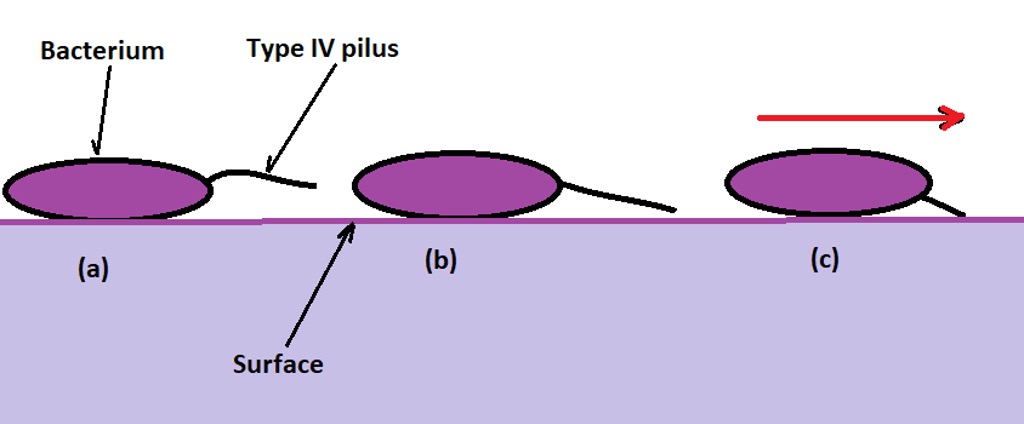
Motility - Apart from adhesion, motility is one of the other important functions of type IV pili. Because of the ability of the pili to retract, they make it possible for the bacterial cell to move along surfaces through a process known as twitching motility. In general, this type of motility takes place through three main stages that include extension, tethering, and retraction.
Following the extension of type IV pili, they adhere/attach to a surface (tethering stage) followed by retraction. This allows the cell to glide over the surface and move in a given direction. This cycle (extension and retraction of type IV pili) has been shown to occur at the rate of about 0.5um per second.
A diagrammatic representation of twitching motility:
(a) the pilus on the surface of the bacteria extending
(b) the extending pilus comes in contact with and attaches/adheres to the surface
(c) the pilus retracts pulling the bacterium in the direction of the arrow (red arrow)
* A single bacterium may have several type IV pili on its surface extending and retracting independent of each other.
Biofilm formation - A biofilm refers to the aggregation of microorganisms which allows them to overcome stressful conditions.
While type IV pili are involved in motility, allowing the cells to move to given sites, they have also been shown to play an important role in the formation of microcolonies and consequently biofilm maturation.
Here, they not only adhere to surfaces and cells, but also promote the close proximity between cells during biofilm formation.
Some of the other functions associated with type IV pili include:
- Electrical conductivity
- Adhesion of bacterial cells to eukaryotic cells (of a host)
- Protein production
Type V Pili
Type V pili is a unique type of pili found in bacteria species of the class Bacteroidia. While the mechanism through which this structure is assembled is not clearly understood, researchers have suggested that it involves protease-mediated polymerization.
Like Type IV pili, this pilus is also involved in adhesion and biofilm formation.
Fimbriae
Also known as "attachment pili", fimbriae are shorter compared to pili and numerous in number (ranging from 100 to 600 filaments per cell).
Depending on the type of bacteria, fimbriae may be located at the poles of the cell or evenly distributed over the surface of the bacterial cell. Because they are shorter, fimbriae are stiffer compared to pili.
There are several types of fimbriae which include:
Type I Fimbriae
Type I fimbriae can be found on the surface of many Gram-negative (E.g., E. coli) where it is involved in adhesion. They range between 1 and 2 um in length and about 7nm in diameter (width). Like all the other fimbriae, type 1 fimbriae are very stiff and do not bend much.
Moreover, they have been shown to be composite structures consisting of a shorter and thin tip fibrillin located at the distal end of the rod. As is the case with pili, type I fimbriae are also characterized by a helical orientation. In this case, the structure has a right-handed helical orientation consisting of 27 FimA subunits with about eight (8) helical turns.
* Type I fimbriae are also known as mannose-sensitive or somatic fimbriae
Functions of Type I Fimbriae
Type I fimbriae are some of the most common structures in the family Enterobacteriaceae. Here, they are involved in the adherence of the bacteria to the cells of the host. However, this binding has been shown to be specific to glycoproteins that consist of one or N-linked high mannose structures.
For this reason, type I fimbriae are assumed to promote infection of the lower urinary tract and some mucosal surfaces.
Type III Fimbriae
Type III fimbriae are thin fimbriae ranging about 5nm in diameter and between 0.5 and 2um in length. Like type II fimbriae, type III fimbriae are especially common among members of the family Enterobacteriaceae and Klebsiella spp.
Like the other fimbriae, assembly of type III fimbriae occurs through the chaperone/usher pathway.
Here, the subunits/building blocks are first transported to the periplasm through the general secretory pathway where a chaperone encoded by the gene mrkB promotes the development and assembly of the fimbriae.
* Like pili and other fimbriae, the subunits (MrKA) of Type III fimbriae are polymerized in a helical orientation.
* Beta strands in the C-terminal region of the subunits provide structural support to the appendage.
Functions of Type III Fimbriae
Type III fimbriae play an important role in adhesion of bacteria to abiotic surfaces as well as the formation of biofilm. For bacteria like K. pneumoniae, attachment to surfaces (e.g., in catheters, etc.) results in aggregation followed by biofilm formation.
This is beneficial for the bacteria in that it promotes pathogenesis by promoting antibiotic resistance among some species of bacteria.
Curli Fimbriae
Curli is another type of fimbriae found in Gram-negative bacteria like Escherichia and Salmonella species. In E. coli, biogenesis of the fimbriae has been associated with at least six (6) proteins which are themselves encoded by csgBA and csgDEFG operons.
Like the other fimbriae and pili, Curli fimbriae also extend from the outer membrane.
Functions of Curli Fimbriae
Curli fimbriae are primarily involved in biofilm formation. For instance, using these fimbria, studies have shown the bacterium Salmonella enteritis to adhere to surfaces like stainless steel and the subsequent formation of biofilm (which contaminate food material).
Differences between Pili and Fimbriae
Both pili and fimbriae are important filaments (consisting of proteins) that originate from the membrane and extend outwards. As such, they are visible under the electron microscope. Because they are antigenic, the two filamentous appendages can also evoke an immune response in vivo.
While they have a number of similarities, the two also have several differences that include:
Size - As mentioned, although they are both shorter and generally smaller compared to flagella, pili are longer than fimbriae with a hair-like appearance. Fimbriae, on the other hand, are shorter with a bristle-like appearance.
Bacteria - Although some Gram-positive bacteria have been shown to possess pili, these structures are commonly found in Gram-negative bacteria. Fimbriae, on the other hand, can be found in both Gram-positive and Gram-negative bacteria where they are involved in adhesion and biofilm formation.
Distribution - Unlike pili, fimbriae are numerous in number and tend to be evenly distributed on the surface of bacterial cells. Whereas a single bacterial cell may contain between 200 and 400 fimbriae on its surface, the number of pili may range from less than 5 to about 10 in total.
Function - While fimbriae are primarily involved in attachment, which promotes biofilm formation, pili are involved in attachment, motility as well as gene transfer from one bacterial cell to another.
Fimbriae have a more solid structure (because they are primarily involved in attachment) while pili, especially sex pili, have a hollow tubular structure that allows for genetic material to be transferred from one cell to another.
Return to Prokaryotes vs Eukaryotes
Return to Unicellular Organisms
Return from Pili and Fimbriae to MicroscopeMaster home
References
Caitlin N Murphy and Steven Clegg. (2012). Klebsiella pneumoniae and type 3 fimbriae: nosocomial infection, regulation and biofilm formation.
Manuela K. Hospenthal, Tiago R. D. Costa and Gabriel Waksman. (2017). A comprehensive guide to pilus biogenesis in Gram-negative bacteria.
Michelle M. Barnhart and Matthew R. Chapman. (2010). Curli Biogenesis and Function.
Stefan David Knight and Julie Bouckaert. (2009). Structure, Function, and Assembly of Type 1 Fimbriae.
Stephen Melville and Lisa Craig. (2013). Type IV Pili in Gram-Positive Bacteria.
Links
https://www.researchgate.net/publication/11238653_Type_IV_Pili_and_Twitching_Motility
Find out how to advertise on MicroscopeMaster!


