Vibrio Bacteria Overview
Examples, Shape, Structure and Infection -
** Vulnificus, Cholerae and Parahaemolyticus
Overview/Introduction to Vibrio Bacteria
With over 100 species, Vibrio bacteria is a genus that is Gram-negative and are widely distributed in aquatic environments (including coastal waters and estuarine, etc). In these environments, the majority of species are associated with a variety of marine organisms including different types of fish and crustaceans , etc (in these organisms, vibrios either exist as commensals or parasites).
Of the 100+ species already identified, only 12 have been associated with human diseases/infections with Vibrio cholerae being the most common pathogen. Aside from causing human diseases/infections, a good number of human pathogens are also responsible for infections in a number of marine organisms.
Examples of species that belong to the genus Vibrio include:
- V. cholerae,
- V. parahaemolyticus,
- V. vulnificus
- V. fluvialis
- V. furnissii
- V. hollisae
Brief History of Cholera and Vibrio bacteria
Between the early and mid 19th Century, severe cholera outbreaks were reported in different parts of the world from India in 1817 to parts of Europe and America in the 1850s. During this period, it was widely believed that the epidemic was the result of poisonous vapor along with particles from decomposing matter.
In the mid 19th Century, however, research studies established that the infections were a result of a microorganism that would later come to be known as Vibrio cholerae.
In 1854, John Snow, regarded as one of the pioneers of modern epidemiology, examined various water samples, and noticed the presence of white flocculent particles in areas with high cases of cholera associated deaths. Although he was able to show that the spread of the disease (cholera) was related to water consumption, he could not identify the organism.
In the same year (1954), by investigating the bodies of patients who died of the disease in Florence, Filippo Pacini, an anatomist, identified tiny elements that he named vibrions.
By inspecting the feces and intestinal mucosa of patients who died, Pacini noticed that all the people who died of the disease were infected by these elements which led him to conclude that it was these vibrions, rather than poisonous vapor, that caused the disease.
However, because he had not conducted any experiment to show evidence of the relationship between the vibrion and the disease, his views were rejected by the scientific community which held that the disease was the result of bad air.
In the 1880s, based on the works of Louis Pasteur, Robert Koch, a German scientist developed various techniques for the purposes of cultivating and studying characteristics of various microorganisms. These techniques allowed him to isolate and culture the organism from the internal mucosa of patients who died of the disease and even identify their general morphology.
Given that Koch only isolated the organism from the bodies of patients who died of the disease, he concluded that this particular microorganism was responsible for the disease.
Through additional experiments, Koch also noticed that the organism multiplied in moist, soiled linen as well as damp earth but was unable to reproduce the disease in animals. Despite providing significant evidence to show the correlation between the organism and the disease, his findings were widely rejected in various parts of Europe.
Classification
Kingdom: Bacteria - As bacteria, members of the genus Vibrio are characterized by a simple cell structure that lacks membrane-bound organelles (prokaryotic). They are also unicellular organisms with a cell wall (consists of a peptidoglycan layer) covering the cell.
Phylum: Proteobacteria - Members of the Phylum Proteobacteria are Gram-negative. As is the vase with Vibrio bacteria, some of the organisms in this group are free-living in the environment while others are pathogenic and thus capable of causing diseases in human beings and animals.
Class: Gammaproteobacteria - The class Gammaproteobacteria is a large and diverse group that consists of Gram-negative bacteria. They are unicellular organisms with the majority of the species having a rod-like appearance.
Order: Vibrionales - Vibrionales is an order that consists of Gram-negative bacteria that are characterized by a straight or curved rod-like morphology. They also move by means of polar flagella (located at one end of the organism).
Family: Vibrionaceae - Vibrionaceae is a family of the order Vibrionales that consists of Gram-negative bacteria commonly found in aquatic (freshwater and marine) environments where they move by means of a flagellum. The majority of these species are also facultative anaerobes that obtain their energy through fermentation.
Genus: Vibrio - Characteristics of the genus Vibrio are discussed below in detail.
Ecology and Distribution
Members of the genus Vibrio are widely distributed in major oceans and various freshwater bodies across the world. According to studies, they make up between 0.5 and 5 percent of the total bacteria community in major oceans across the world. This is a significant number considering that the ocean consists of over two (2) million species of bacteria.
Although they can be found in estuaries (blackish water bodies connected to one or more rivers as well as the open sea), these bacteria are indigenous in marine environments.
In the marine environment, the general growth, and distribution of Vibrio bacteria are dependent on various environmental factors. For instance, the population of Vibrio species (e.g. V. vulnificus and V. parahaemolyticus etc) has been shown to increase at temperatures over 20 degrees C with the population peaking between 20 and 30 degrees C.
Changes in temperature have also been shown to directly influence the risk of infection for human beings. In the United States, for instance, over 75 percent of Vibrio related infections occur at water temperatures over 20 degrees C (between the months of May and October).
Members of the genus Vibrio found in freshwater, particularly V. cholerae are responsible for various outbreaks especially in rural areas. While these diseases/infections have been reported in all regions across the world, they are more common in rural and third world countries: In these areas, poor hygiene is one of the factors that significantly contribute to the spread of cholera.
During harsh environmental conditions, some of the species in temperate marine areas have been found to live in sediments. This habitat has been shown to be particularly ideal under such circumstances (harsh conditions) given that it contains high amounts of organic material that can support the bacteria for a given period of time.
On various surfaces, on the other hand, Vibrio bacteria survive by forming biofilms that allow them to live and survive in different environments including water and other moist surfaces.
* As commensals or pathogens, Vibrio bacteria can be found living within the host (e.g. in the intestines) or attached to the surface of the host.
Morphological Characteristics of Vibrio Bacteria
Like many other bacteria, members of the genus Vibrio are small in size, ranging from 0.5 to 0.8 um in diameter and 1.4 to 2.6 um in length. However, in a culture, they can form colonies that are between 2 and 3 mm in diameter.
They are characterized by a straight or curved (comma-shaped) rod-like shape as well as a single polar flagellum used for movement. Although some of the Vibrions have a straight, rod-like shape, members of this group, e.g. V. cholerae, are curved, one of the characteristics used to classify these bacteria (classification based on shape/morphology).
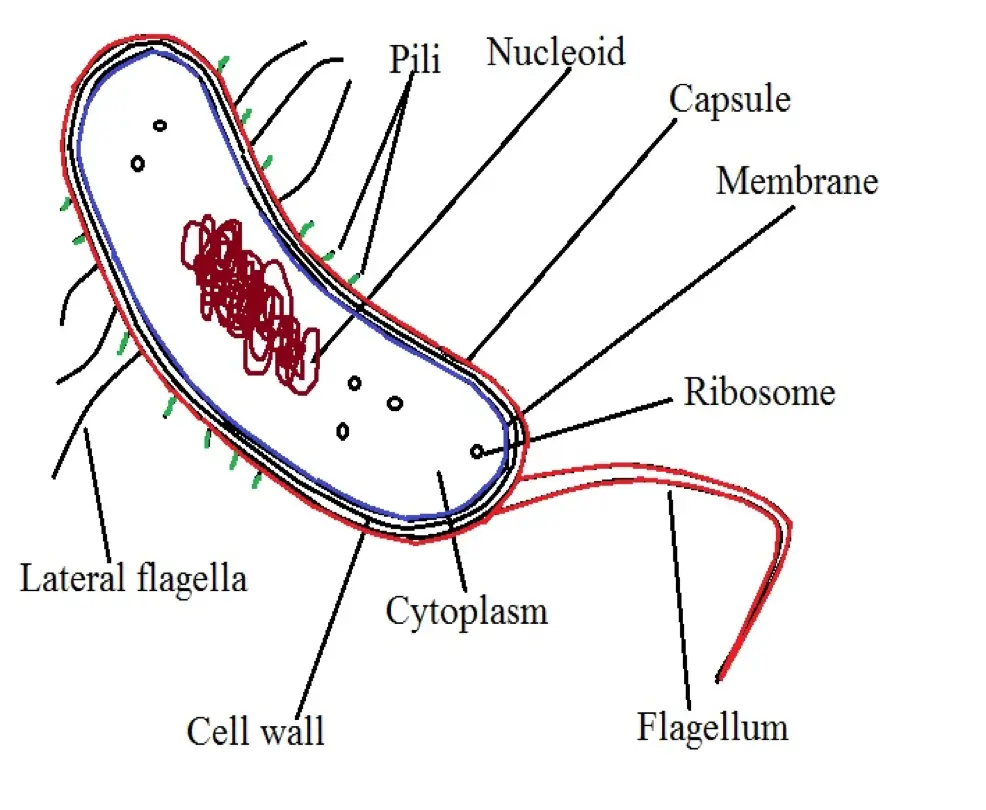
The following is a diagrammatic representation of a Vibrio bacteria cell:
The cell wall of Vibrio bacteria, like that of many other bacteria, consists of a peptidoglycan layer. The glycan chains, which make up the peptidoglycan layer, encircle the cell body in width (around the cell body like a telephone cord).
It's worth noting that in some of the species, individual chains are not long enough to completely encircle the entire circumference of the cell body. For this reason, some of the chains may appear like short fragments that only run a certain distance around the circumference of the cell.
Unlike most coccoid and bacillus, Vibrio species are highly mobile and use a flagellum to move from one location to another. As already mentioned, members of this group are commonly found in aquatic environments. Here, then, having this structure is particularly important as it allows them to swim freely.
Generally, the number and type of flagella produced are largely dependent on the species. While the majority of species have monotrichous polar flagella (one larger, polar flagella, and several lateral flagella), some of the species use lophotrichous or peritrichous for movement.
* Movement of Vibrio bacteria is made possible by the rotating or propelling mechanism of the flagella.
The structure of a flagellum consists of three main parts.
These include:
- The basal body
- The hook
- The filament
In liquid environments (aquatic environments), such species as V. parahaemolyticus only have a single polar flagellum located on one end of their cell body. The flagellum is covered by a sheath covering that extends from the cell membrane of the organism.
In this environment, a single flagellum is sufficient for movement. However, in an environment with a more viscous liquid or a semi-solid, additional flagella, known as lateral flagella, are produced to allow the organism to move more effectively. These structures are more useful for adhesion/attachment which in turn allows the bacteria to move.
* Apart from flagella, some species produce a protein structure known as MAM7 (multivalent adhesion molecule) that allows the bacteria to adhere to the cell of a host in the event of an infection.
While they lack membrane-bound organelles as prokaryotes, Vibrio bacteria have a number of important organelles and structure that include:
- Nucleoid
- Riboplasm
- Cytoplasm
- Pili
- Cell membrane
- Cell capsule
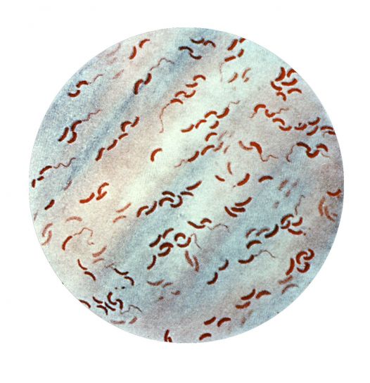
* As Gram-negative bacteria, Vibrio have a considerably thin peptidoglycan layer compared to that found in Gram-positive bacteria. For this reason, they do not retain the primary stain and thus take up the color of the counterstain. When observed under the microscope, they will appear as curved pink elements.
* To study the morphological characteristics of Vibrio bacteria, direct dark-field microscopy is recommended - Using this technique, it's possible to identify motility characteristics of these organisms.
Read more about Gram negative and Gram positive bacteria
Take a look at other Microscopy Imaging Techniques
Nutrition
Like many other bacteria, Vibrio bacteria are heterotrophic and thus incapable of producing their own food. For this reason, they depend on various organic materials as a source of energy.
For different species, metabolism has been shown to be oxidative (where oxygen is used to break down and obtain energy from carbohydrates) or fermentative (degradation of organic matter anaerobically to produce energy). However, fermentative metabolism is the most common mode of metabolism for the majority of species.
* Given that the majority of these species are not fastidious, they can survive on simple carbon sources like glucose as their source of energy.
Although the majority of species exist as free-living organisms in marine and freshwater environments, some of the species (e.g. Vibrio cholerae) are pathogenic and cause diseases in human beings (other species cause diseases in both human beings and other animals). These species can be found in contaminated water sources or even fruits/food.
Bioluminescence
Bioluminescence refers to the ability of some organisms to emit light. Vibrio fischeri, a member of the genus Vibrio is one of the few bacteria that produce light.
Light is produced through a reaction in which an oxidative enzyme known as luciferase (consisting of LuxA and LuxB) transforms FMNH2 (a form of flavin mononucleotide), oxygen, an aliphatic aldehyde to Flavin mononucleotide, water, and aliphatic acid. Generally, this process is regulated by the LuxR-LuxI quorum-sensing system.
Pathogenesis and Pathophysiology
For the most part, Vibrio infections occur when individuals/animals ingest contaminated foods or water sources. This is one of the main reasons why cholera outbreaks are common in areas/regions where people do not practice good hygiene.
Once the organism is ingested, the majority of them are destroyed by gastric acid in the stomach. Surviving organisms successfully colonize this region by adhering to the intestinal mucosa.
Adhesion of the bacteria may occur through association of the organism with the mucus/gel in the intestine. The bacteria adheres to the epithelial wall. To adhere to the epithelial cells, these bacteria have been shown to produce such protein structures as a multivalent adhesion molecule.
Following adhesion, Vibrios may cause the disease (e.g. cholera) by releasing toxins (e.g. protein exotoxin) that ultimately result in watery diarrhea. By releasing the toxin, which consists of an A subunit that is attached to five B subunits, the B subunit binds to a region on the epithelial cell which in turn allows the A subunit to enter the cell.
Within the cell, this subunit activates adenylate cyclase thus raising the intracellular cyclic AMP. As a result, chlorine secretion is increased which ultimately causes watery diarrhea.
* According to statistics, there are between 3 and 5 million cases of cholera annually with as many as 100,00 to 120,000 deaths.
* As mentioned, 12 Vibrio species are responsible for infections (gastrointestinal illnesses) in human beings. The illnesses caused by Vibrio bacteria are known as vibriosis.
Apart from contaminated water and fruits etc, Vibrio bacteria can also be transmitted by various marine organisms including shellfish, mollusks, and crustaceans among others.
Here, the bacteria are often transmitted to human beings when they consume fish and shellfish etc that have not been fully cooked. Failure to keep these marine organisms (fish, etc) cold once they are harvested causes them to continue dividing and multiplying thus increasing the risk.
While V. cholerae is largely transmitted through contaminated water, studies have shown V. parahaemolyticus and V. vulnificus to be some of the most common Vibrio species that cause food-borne infections.
· Mollusks are responsible for about 45 percent of food-borne infections
· Fish are responsible for about 38 percent of these outbreaks
· Crustaceans contribute about 16.0 percent of the outbreaks
* Properly handling food (especially marine organisms) and maintaining good hygiene is one of the best means of controlling the spread of these bacteria.
Reproduction
Whether in freshwater, human host, or marine environments, Vibrio bacteria use binary fission as their primary mode of reproduction. Under favorable environmental conditions, the organism divides to form two similar daughter cells that have the capacity to grow to the size of the original/parent cell allowing the cycle to continue.
This process starts with the elongation of the cell body as the DNA is replicated in order to produce two copies to be shared between the two daughter cells. The cell wall along with the plasma membrane of the bacteria then invaginates near the middle as cross walls start forming to separate the two cells.
Ultimately, the two cells divide/separate into two to form two similar daughter cells. Under conducive environmental conditions (nutrition and optimal temperature etc), the two new daughter cells grow and the process is repeated.
Return to Bacteria shape, Size and Arrangement
Return from Vibrio Bacteria to MicroscopeMaster home
References
D. Lippi and E. Gotuzzo. (2013). The greatest steps towards the discovery of Vibrio cholerae.
Jason B Harris, Regina C LaRocque, Firdausi Qadri, Edward T Ryan, Stephen B Calderwood. (2012). Cholera.
Jeffrey L. Bose et al. (2007). Bioluminescence in Vibrio fischeri is controlled by the redox‐responsive regulator ArcA.
Mylea A. Echazarreta and Karl E. Klose. (2019). Vibrio Flagellar Synthesis.
Stephen Cooper. (2001). Helical growth and the curved shape of Vibrio cholerae.
Links
https://www.sigmaaldrich.com/technical-documents/articles/microbiology-focus/vibrio-detection.html
https://www.fda.gov/food/laboratory-methods-food/bam-chapter-9-vibrio
Find out how to advertise on MicroscopeMaster!


