Eubacteria
Examples, Characteristics, Vs Archaebacteria
Definition
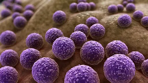
Also referred to as "true bacteria" in some books, Eubacteria is a domain consisting of all the common groups of bacteria. As such, it's made up of all species that fall within the Bacteria domain.
While all groups within this domain are prokaryotes, they display high diversity in their general morphologies, metabolism, and habitats. This has made it possible to classify and describe different types of bacteria in nature.
Various groups exist as parasites and are responsible for animal and plant diseases. However, some are free-living with some being beneficial to man.
Some of the groups of bacteria that fall under the domain Eubacteria include:
- Proteobacteria
- Cyanobacteria
- Gram-positive bacteria
- Spirochetes
- Chlamydias
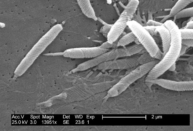
Proteobacteria
Proteobacteria represent the largest and most diverse group among prokaryotes. As such, the division includes various parasitic forms (e.g. Salmonella, Helicobacter, and Escherichia etc) as well as free-living forms such as those involved in nitrogen fixation.
All members of the group are Gram-negative and thus do not retain the primary stain during Gram staining. Based on rRNA sequences, this division is divided into six major groups that include; Alpha, Beta, Delta, Epsilon, Gamma, and Zeta proteobacteria.
Based on metabolism, the division is also divided into three main subgroups that include Purple bacteria (photoautotroph and photoheterotroph that contain chlorophylls), Chemoautotrophic proteobacteria (consisting of free-living and symbiotic members), and Chemoheterotrophic proteobacteria (also known as enteric bacteria).
Cyanobacteria
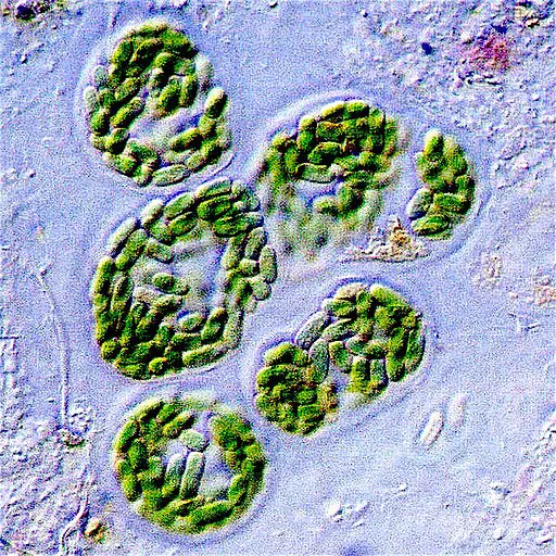
Also known as blue-green algae, Cyanobacteria is a class of Eubacteria consisting of photosynthetic microbes (commonly referred to as photosynthetic prokaryotes). These prokaryotes are ubiquitous in nature and can, therefore, be found in such environments as ponds, rivers, lakes, desert soil, and hot springs among others.
In these environments, some species are free-living (given that they are capable of photosynthesis) or may form symbiotic relationships with fungi to form lichens.
Some of the other characteristics associated with Cyanobacteria include a thick and gelatinous cell wall; gliding among motile species and diverse morphological characteristics. They may exist as single-cells, colonies or as multicellular organisms where different cells perform different functions.
See Also: Schizophyta
Sprirochetes
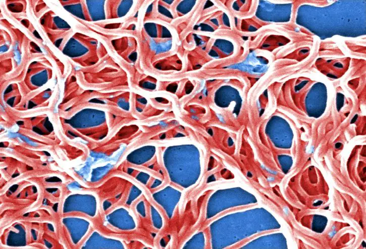
As the name suggests, spirochetes are characterized by a spiral morphology. This architecture is the result of a flexible peptidoglycan cell wall covered by axial fibrils.
Although spirochetes are Gram-negative, they tend to stain poorly which makes it challenging to visualize using routine microscopy. In nature, Spirochetes can be found in water bodies, decaying organic matter, animals, plants and soil, etc.
Like many other groups, the group consists of both free-living and pathogenic organisms that have been shown to cause diseases in human beings and animals (e.g. Borrelia burgdorferi and Treponema pallidum).
Some of the other characteristics associated with spirochetes include:
- Covered by a bilayered membrane
- Some species form a hyaluronic acid slime layer
- Movement is made possible by the presence of an internal flagellar filament (causes spinning and flexing about a long axis)
Chlamydias
Chlamydias is a group of organisms belonging to the family Chlamydiaceae. They are obligate intracellular parasites of animals and are the most common causes of non-gonococcal urethritis in the United States. Chlamydia trachomatis, on the other hand, is the most common cause of blindness across the globe.
While Chlamydias species are grouped as gram-negative, their peptidoglycan is not easily detectable. Some of the other characteristics of Chlamydias include; they are immotile and are dimorphic in nature.
Gram-positive Bacteria
A majority of Eubacteria are Gram-positive. Whereas some of the species in this group are capable of photosynthesis (photosynthetic Eubacteria), a majority are chemoheterotrophs and thus obtain nutrients from their surroundings.
Being Gram-positive bacteria, these species retain the primary color (Crystal violet dye) during Gram staining and thus appear violet in color when viewed under the microscope. This is because they contain a thick peptidoglycan layer compared to Gram-negative species.
They vary in morphology from cocci species that are spherical in shape (e.g. Staphylococcus) to rod-like species (bacilli) such as Listeria.
Depending on the specie, Gram-positive bacteria can be found in different types of aquatic (marine and freshwater) and terrestrial environments. In the environment, various species (e.g. Mycoplasmas and Actinomycetes) have been shown to form endospores.
While many of the species are parasitic and capable of causing diseases in animals, some of the species (e.g. Actinomycetes) are beneficial in that they produce medically relevant compounds such as antibiotics.
Major Characteristics of Eubacteria
Metabolism
Because of the diversity of species classified under the domain Eubacteria and the fact that they are found in a wide range of habitats, they have been shown to display a higher diversity in their energetic sources and metabolic pathways when compared to eukaryotes.
In the absence of organic matter, many species (described as chemoautotrophic eubacteria) have been shown to obtain their energy through the oxidization of mineral compounds.
A good example of this is bacteria species that oxidize nitrates or methane. In the process, they also play an important role in converting various material into useable compounds.
Using such compounds as ammonia, nitrifying bacteria produce nitrates that are used by plants to make proteins and amino acids etc.
Some of the main nutritional modes of different species in the domain Eubacteria include:
· Autotrophic - Produce their own food through photosynthetic processes
· Heterotrophic - Obtain nutrients from their environment (because they are unable to synthesize their own food)
· Strictly or facultatively aerobic - Survive in the presence of oxygen (strict aerobes) or can switch to anaerobic respiration in the absence of oxygen (facultative anaerobes)
· Strictly or facultatively anaerobic - Survive in the absence of oxygen (strict anaerobes) or can survive with or without oxygen (facultative anaerobes)
For Eubacteria, diversity in the modes of metabolism is also due to the wide variety of chemical compounds that serve as substrates as well as the ability to produce energy in different environmental conditions.
Whereas various photosynthetic forms require oxygen for photosynthesis, others can depend on fermentation processes for energy and only grow well in the absence of oxygen.
In the food chain, they play an important in various environments not only as prey of various unicellular eukaryotes, but also as producers that support various metazoans and decomposers that break down dead material that is then re-used by plants.
Cell Structure
All eubacteria species have a cell wall consisting of peptidoglycan. However, according to various studies, species that lack this structure are those that have lost it secondarily. Peptidoglycan is made up of glycosaminoglycan which in turn consists of alternating residues of D-glucosamine and muramic acid.
During peptidoglycan synthesis, the carboxyl group of the muramic acid is typically substituted by a peptide consisting of L- and D-amino acid residues.
Depending on the organism, these peptide units may be cross-linked through peptide bonds to produce a giant macromolecule that contributes to the cell wall.
In Gram-negative species, the macromolecule occurs as a monomolecular layer located between the inner and outer membrane. In Gram-positive bacteria, it appears as a multimolecular layer covalently or non-covalently bound to such compounds as teichoic acid or neutral polysaccharides.
* Typically, bacteria that lack a cell wall is given the suffix "plasma". Some examples include Ureaplasma and Mycoplasma. These characteristics allow them to remain resistant to various antibiotics that function by targeting the cell wall structure.
* In Gram-negative bacteria, the peptidoglycan layer makes up about 80 percent of the cell wall.
Reproduction
While some species have been shown to exchange genetic material through cell contact, asexual reproduction is the main mode of reproduction in eubacteria.
Some of the main methods of asexual reproduction in eubacteria include:
Binary Fission
This is the most common mode of reproduction in Eubacteria during favorable environmental conditions. During binary fission, genetic material of the organism (nucleoid) divides without mitosis. This occurs through DNA replication, a process that produces two strands of DNA.
DNA replication, which is bidirectional, originates from a given point, known as the point of origin, producing two circular DNA molecules.
The cell divides into two when a peripheral ring of the membrane invaginates and grows towards the central part of the cell thereby forming a double-membranous septum. Cell wall materials are then deposited between the two membranes causing the two cells to separate completely.
Endospore Formation
For the most part, endospore formation occurs during unfavorable environmental conditions and is common among Gram-positive bacteria and cyanobacteria.
During unfavorable environmental conditions (extreme temperature, presence of toxins or nutrient deficiency etc) these organisms produce a thick wall to surround the cell to form the endospore.
Before the cell is encased in the thick cell wall, the chromosome and parts of the cytoplasm are also dehydrated. The cytoplasm and cell wall also deteriorate giving way to the thick wall that ultimately covers the cell. This makes the organism resistant to unfavorable conditions and allows it to survive for a long period of time (up to several decades).
The thick wall covering the endospore consists of four main parts:
- Exosporium - Loose outer covering
- Cortex - Thick layer consisting of mucopeptides and carbohydrates
- Core wall - Also known as the inner cortex, the core wall is largely composed of proteins
- Spore-coat - Consists of disulfide-rich proteins and resembles keratin
* Under favorable conditions, the protoplast swells up when it absorbs water allowing dipicolinic acid to leak out. This activates and causes the protoplast to break the spore cover. Ultimately, a bacterial cell emerges from the spore and continues developing and reproducing under favorable conditions.
Some of the other common forms of asexual reproduction in Eubacteria include:
· Fragmentation - The organism breaks into fragments after growing to a given length as seen in cyanobacteria
· Budding - The organism produces buds that grow and detach to develop into new individuals
· Conidia formation - Common in mycelial bacteria and involves the development of new individuals from conidia (rounded structures cut from parts of the bacteria)
* Sexual reproduction in Eubacteria involves an exchange of genetic material through conjugation, transduction, and transformation.
Eubacteria Vs Archaebacteria
Like Eubacteria, archaebacteria is a branch of prokaryotic evolution. However, unlike Eubacteria, studies have shown them to be simpler and much ancient.
For this reason, their cellular make-up is noticeably different from that of Eubacteria. For instance, members of this group (Archaebacteria) lack peptidoglycan and contain RNA polymerase and ribosomal proteins more similar to those of eukaryotes than eubacteria.
Some examples of Archaebacteria including:
- Ferroplasma
- Halobacterium
- Pyrobaculum
- Lokiarchaeum
- Thermoproteus
* Some of the species possess a pseudopeptidoglycan. In these species, the N-acetylmuramic acid is replaced by N-acetyltalosamine uronic acid.
For a majority of the species, the cell wall consists of a surface layer known as "S-Layer". It's made up of proteins/glycoprotein arranged in a hexagonal pattern and assemble into lattices that range between 5 and 25nm in thickness.
Despite the difference in structure from that of Eubacteria, this composition serves to provide mechanical support to the cell in addition to their role in preventing osmotic lysis.
Apart from the differences in cell wall composition, differences between Eubacteria and archaebacteria are also evident in their cell membrane. Among archaebacteria species, hydrophobic lipid tails are bound to glycerol through ether linkages instead of ester linkages present in Eubacteria species.
On the other hand, the fatty acid tails common in Eubacteria are replaced by repeating units of five carbon that are linked in a manner allowing them to form tails that are noticeably longer than fatty acid tails.
In cases where this produces a lipid monolayer, this provides an advantage to hyperthermophiles given that the structure prevents the membrane from peeling into two.
Members of the domain (Archaea) are divided into two phyla. These include:
· Euryarchaeota - This group consists of extremophiles capable of surviving highly unfavorable conditions. A good example of these are methanogens commonly found in environmental devoid of oxygen and extreme halophiles that can be found in environments with high salt concentrations.
· Crenarchaeota - Organisms classified under this phylum are hyperthermophiles, normally found in hot aquatic environments (springs and marine environments)
Some of the other characteristics of Archaebacteria include:
- Vary in shape morphology (e.g. rod-shaped, spherical, flattened)
- Contain introns (non-coding sections of RNA)
- Reproduce asexually through binary fission, fragmentation and budding
- Do not exhibit glycolysis
See also: Fusobacteria
How do Bacteria cause Disease?
Return to learning more about Bacteria size, shape etc.
Return from Eubacteria to MicroscopeMaster home
References
Chia Y. Lee and David Cue. (2013). Introduction to Bacteriology.
Guillaume Lecointre and Hervé Le Guyader. (2007). The Tree of Life: A Phylogenetic Classification.
Jones and Bartlett Publishers. Cell Structures and Function in the Bacteria and Archaea.
Larry L. Barton. (2005). Cell Wall Structure. In: Structural and Functional Relationships in Prokaryotes. Springer, New York, NY
Links
https://www.sciencedirect.com/topics/earth-and-planetary-sciences/archaebacteria
https://accessmedicine.mhmedical.com/content.aspx?bookid=1020§ionid=56968779
Find out how to advertise on MicroscopeMaster!
