Somatic Cells
Types, Location, Process of Production, Vs Germ Cells
Types of Somatic Cells
|
Somatic cells are all cells of the body apart from gamete (sperm cells and egg cells). As such, they include cells that make up different parts of the body including liver cells, skin cells, and bone cells among others. Mature somatic cells are highly specialized and therefore perform very specific functions. |
* The word somatic is derived from the Greek word "Soma" which means body.
The following are some of the most common types:
Epithelial Cells
Epithelial cells include different types of cells that make up the epithelial tissues. As such, they line different organs and parts of the body including the skin, the blood vessels, the digestive tract among other body cavities and hollow organs.
Depending on the type of cells, epithelial cells perform a variety of functions ranging from secretion to excretion.
Some examples of epithelial cells include:
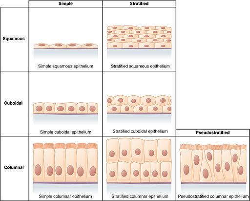
Squamous cells - These cells are thin and flattened. They are also large and are characterized by a small, round central nucleus as well as abundant cytoplasm. As compared to the nuclei of other cells, the nuclei of these cells also appear flattened and elliptical.
These cells can be found lining different parts of the body including the lung air sacs, the epidermis, capillaries, distal part of the urethra in men as well as the urethra in women. Because they are exposed to the external environment like in the epidermis and various substances and chemical compounds inside the body, these cells are lost and replaced regularly.
Cuboidal cells - Cuboidal epithelial cells are characterized by a cube-like morphology. In addition to the typical cell organelles, these cells are also characterized by the presence of secretory vesicles and microvilli.
As such, they are commonly found in parts of the body associated with high metabolic activities where they are involved in the secretion and exchange of various substances. They can be found in various ducts, tubules (e.g. kidney tubules), and the neural retina, etc.
Columnar cells - Unlike cuboidal cells, columnar cells, as the name suggests, are elongated cells and are therefore taller than they are wide. Some of these cells, particularly simple columnar cells, are ciliated and are involved in secretion and absorption.
Columnar cells can be found lining the fallopian tubes, parts of the respiratory tract, as well as digestive tract (particularly the stomach and anal canal).
* Transitional epithelial cells include cells that are capable of changing shape.
Cells of the Connective Tissue
Connective tissue is one of the most abundant and widely distributed tissues of the body. It serves a number of important functions including providing protection, binding functions, as well as support. As such, it consists of several types of cells that are specialized for these functions.
In general, cells of the connective tissue are divided into two main groups that include:
Resident Cells
These are cells of the connective tissue that are fixed and therefore do not migrate from the connective tissues.
These cells include:
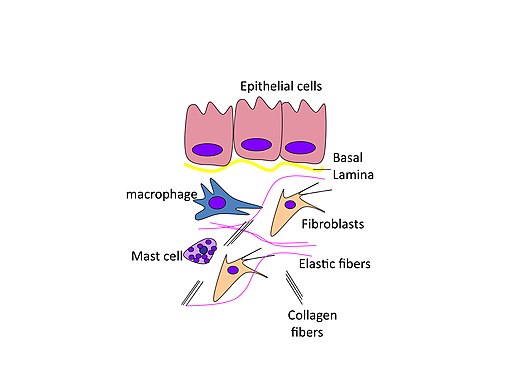
Fibroblasts - Fibroblasts are some of the most common cells of connective tissue. They are characterized by a spindle-shaped (or stellate shaped) morphology as well as a flattened/ovoid nucleus.
Being some of the most abundant cells of connective tissue, fibroblasts can be found in the interstitial spaces of various organs.
As resident cells of connective tissue, fibroblasts remain fixed at given regions of organs (liver, kidneys, lungs, etc). They have been shown to remain quiescent until they are activated and are involved in the production of various products including laminins, fibronectin, collagen, and prostaglandins among others.
In doing so, fibroblasts play an important role in reorganizing the extracellular matrix and wound healing.
Macrophages - Resident macrophages originate from erythromyeloid progenitors (which originate from the yolk sac). Being resident cells, macrophages reside in the tissue where they constitute the mononuclear phagocyte cellular system.
In the body, macrophages respond to pathogenic invasions where they are involved in the ingestion of invading organisms as well as damaged cells.
Following the depletion of tissue macrophages, studies have shown there to be an increased level of collagen and hyaluronan. This led to the conclusion that in tissue, these cells also play an important role in ECM (extracellular matrix) homeostasis.
Adipocytes - Also known as fat cells, adipocytes are cells of the connective tissue that originate from the mesenchymal stem cells. In addition to other cell organelles, these cells are characterized by a large fat droplet located near the central part of the cell. This is the part of the cell in which fats (triglycerides) are stored.
With the increase in fat content in the body, the number of these droplets, in brown adipocytes, has been shown to increase within the cell. However, they do not decrease with decreased fat content. In addition to fat storage, adipocytes are also involved in energy generation as well as maintaining body temperature.
Mast cells - Mast cells are commonly found in the mucosal and epithelial tissues. Generally, they are characterized by an oval shape. Mast cells originate from the bone marrow from where they migrate to the connective tissue (particularly the loose connective tissue).
They are also characterized by basophilic granules involved in the production of histamine and heparin. Whereas heparin is a coagulant, histamine is involved in allergic reactions. Following the release of histamine, cell junctions are weakened which allows proteins and cells to enter the connective tissue.
Transient Cells
Unlike resident cells, transient cells of the connective tissue migrate to the connective tissue from the bloodstream in order to get to the affected site.
As such, they include cells of the immune system including:
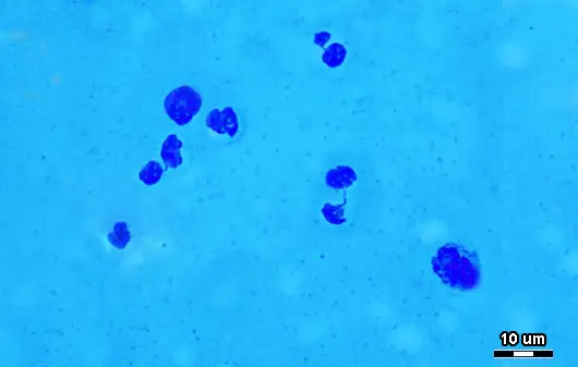
Neutrophils - Granulocytes that make up between 40 and 70 percent of the total leukocyte count. They are characterized by a multilobed nucleus (consisting of between 3 and 5 lobes) and are part of the first line of defense against invading microorganisms and also signal the production of other immune cells.
Eosinophils - Eosinophils are generally characterized by a bilobed nucleus and large cytoplasmic granules. Although they are involved in allergic reactions, they are also involved in fighting off multicellular organisms.
Basophils - Basophils make up between 0.5 and 1 percent of the total leukocyte count and tend to be larger compared to the other granulocytes. As granulocytes, they contain granules that produce enzymes involved in allergic reactions. In addition, they are also involved in blood clotting processes.
Monocytes - Monocytes are large cells that can differentiate to produce macrophages and dendritic cells. In addition to removing damaged cells through phagocytosis and fighting given infections, these cells also play an important role in regulating immunity against invading organisms and foreign substances.
Plasma cells - Also known as B cells, plasma cells are involved in the production of antibodies in response to a given antigen which allows them to be identified and destroyed.
Nerve Cells
Also known as neurons, nerve cells are cells of the nervous system that transmit and process chemical/electrical signals. As such, they may be described as signaling units of the nervous system.
Nerve cells are characterized by three main parts that include the cell body which contains the nucleus and various cell organelles, dendrites that originate from the cell body (receives nerve impulses), and the axon which extends from the cell body (transmits a nerve impulse).
* Generally, nerve cells allow organisms to respond effectively to stimuli. For this to be achieved, the cells first receive information from the environment or other nerve cells, process this information, and ultimately send out information to the effector tissues which allows them to respond appropriately.
* Apart from nerve cells, the other type of cell that belongs to the nervous system is known as neuroglia or glial cells. Glial cells have a radial morphology.
As compared to neurons, glial cells do not produce electrical impulses and therefore are not involved in transmitting information. Rather, they help maintain homeostasis and provide structural support for nerve cells.
Cells of Muscular Tissue
The muscular tissue is an important part of the muscular system that functions by contracting to allow for movement.
This tissue consists of three types of cells that include:
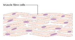
Skeletal cells - skeletal cells or skeletal muscle cells are characterized by an elongated morphology as well as an elastic plasma membrane. In addition, they are striated and contain more than one nucleus.
As well as their ability to contract which contributes to various movements, skeletal muscle cells are also involved in the production of various materials including endocrine, bioactive, and autocrine factors.
Cardiac muscle cells - Unlike skeletal muscle cells, cardiac muscle cells (also known as cardiomyocytes) are characterized by fairly rectangular morphology and a single nucleus. They are also actively involved in contraction movements and thus contain numerous sarcosomes as sources of energy.
The ends of these cells are joined together by intercalated disks, involved in cell-cell communication, which produce long fibers. This is a unique characteristic of cardiac muscle cells.
Smooth muscle cells - These cells are characterized by a spindle shape and generally contain a single nucleus. Like some of the other cells of the muscular system, smooth muscle cells are elastic and therefore allow for the expansion and contraction of various organs (lungs, kidneys, etc).
Location Somatic Cells
As mentioned, somatic cells are highly specialized to perform given functions. As such, they can be found in different parts of the body where they are involved in given functions associated with those parts of the body.
The following are some of the locations in which different types of somatic cells are found:
Cells of Epithelial Tissue
Squamous epithelial cells - Simple squamous epithelial cells play an important role in secreting lubricating substances as well as allowing various substances to pass through.
They are commonly found in regions of the body associated with these functions including blood and lymphatic vessels, the alveoli, as well as serous membrane that line body cavities and the surface of internal organs.
Stratified squamous epithelial cells consist of several layers of flattened cells as well as basal layers of columnar or cuboidal cells. These cells play an important role in providing protection against abrasions and are therefore found in regions of the body that are likely to be exposed to the external environment including the skin, the mouth cavity as well as parts of the body like the vagina.
Cuboidal epithelial cells - These cells are characterized by a cuboid morphology and are generally involved in the secretion and absorption of various substances.
Simple cuboidal epithelial cells (a single layer of cells) can be found in the bronchioles as well as secretory parts of the small glands, surface of the ovaries, the lining of the renal tubules, as well as parts of the thyroid.
Stratified cuboidal epithelial cells can be found lining the surface of excretory ducts such as the sweat glands as well as parts of the kidney tubules.
Columnar epithelial cells - Simple columnar epithelial cells are also involved in absorption and secretion (e.g. secreting mucous). Moreover, they also play an important role in moving mucous.
They are therefore commonly found in the in parts of the digestive system, the fallopian tube in the female reproductive tract, minor ducts, as well as the uterine cervix.
Stratified columnar epithelial cells are commonly found in the conjunctiva as well as some sections of the uterus, pharynx, and the anus etc.
Connective Tissue Cells
The connective tissue originates from the mesoderm and commonly found between other tissues of the body. Some of the functions associated with this tissue include providing structural support, protecting body tissues and organs, transportation, insulation, as well as storage.
Resident Cells
As mentioned, resident cells are generally fixed at given regions and do not migrate.
The following are some of the locations in which these cells can be found:
Fibroblasts - Fibroblasts are the most abundant cells of the connective tissue and are therefore found in all organs and tissues of the body. Based on cultural studies, these cells have been shown to transform and give rise to other cells of the connective tissue under certain conditions.
Macrophages - Macrophages are important cells of the immune system and therefore play an important role of protecting the body against bacteria and other microorganisms. As such, they can be found in many parts of the body including the lungs, spleen, skin, and lymph nodes among other tissues.
Adipocytes - As mentioned, adipocytes are involved in fat storage and maintaining body temperature. As such, they can found in many parts of the world surrounding internal body organs, beneath the skin, in the bone marrow, as well as in the muscular system etc.
Mast cells - Commonly referred to as master regulators of the immune system, mast cells can be found in all body tissues.
Transient Cells
Blood cells - Blood cells include red cells, leukocytes (white blood cells), platelets, and plasma cells. Although white cells can be found in the bloodstream, they can leave circulation and migrate to different parts of the body (tissue) to fight off infections.
Nerve cells - Given that nerve cells are involved in the transmission of impulses, they make up the central nervous system as well as the peripheral nervous system. As such, they extend to all parts of the body.
Cells of Muscular Tissue
As already mentioned, the muscular tissue consists of the cardiac, skeletal, and smooth muscle cells.
These cells are found in different parts of the body where they are involved in different functions:
Cardiac cells - Also known as cardiomyocytes, cardiac cells are the chief cells of the cardiac muscle (heart muscle).
Skeletal cells - Skeletal muscle cells make up the skeletal muscles which attach to the bones through tendons. As such, they can be found in different muscle tissue involved in movement.
Smooth muscle cells - These are a type of muscle cells that lack striations. They are commonly found in the walls of various hollow organs of the body including the intestines, the bladder, and blood vessels, etc.
Processes of Production
Essentially, all cells originate from the three (3) germ layers (ectoderm, mesoderm, and endoderm). While mesodermal cells give rise to cells of the connective tissue, bone, lymph vessels, and the gonads, etc, ectodermal cells produce cells of the central nervous system, the peripheral nervous system, and epidermis among others.
Ectodermal cells produce cells of the respiratory tract, thyroid, urinary bladder, and the pancreas, etc.
Following the development of the embryo, descendants of the stem cells known as progenitor cells located in different parts of the body differentiates to produce different types of cells.
The following are some of the processes involved in the production of different types of somatic cells:
Hematopoietic Progenitor Cells
Hematopoietic progenitor cells are located in the bone marrow and the peripheral blood. Differentiation of these cells results in the production of specialized blood cells including platelets, white cells, as well as red blood cells.
In order to produce mature functional cells, hematopoietic progenitor cells first divide to produce multipotent progenitor cells which include common myeloid progenitor and commonly lymphoid progenitor (also known as myeloid stem cells and lymphoid stem cells respectively).
From this point, the type of cell produced is largely dependent on the needs of the body. In the event of hypoxia, erythropoietin, which is a growth factor, is produced in the kidney and stimulates the production of red cells.
In the presence of this hormone, myeloid stem cells undergo several stages of division to produce mature red blood cells.
Signal molecules like interleukin 3 and 5 as well as granulocytic and agranulocytic colony-stimulating factors influence the production of granulocytes and monocytes from the myeloid stem cells. Unlike red cells, which lack a nucleus, some of the leukocytes are capable of mitosis.
* Myeloid stem cells give rise to a variety of blood cells including platelets, red cells, monocytes, osteocytes, dendritic cells, and granulocytes.
On the other hand, lymphoid stem cells are responsible for the production of B and T lymphocytes, natural killer cells and some dendritic cells.
Neural Progenitor Cells
Another type of progenitor cell in the body is known as neuronal progenitor cells. These cells are located in the pats of the brain including the lateral ventricle as well as the striatum. In adult mice, studies have shown these cells to differentiate and give rise to functional neurons and glial cells.
However, in adult human beings, these cells have only been shown to give rise to glial cells. For instance, in the Subventricular Zone, the neural progenitor cells (also known as B1 cells) divide mitotically to produce quiescent or proliferative B1 cells.
In turn, these cells are capable of asymmetrical division and give rise to B1 cells that can self-renew as well as transient progenitor cells that can give rise to a group of other cells known as C cells.
Endothelial Progenitor Cells
This is another group of stem cells found in the bone marrow. As is the case with the production of blood cells, endothelial progenitor cells are activated through the production of certain cytokines, growth factors as well as cell-activating factors.
These substances may be produced in the event of an injury and influence the mobilization of the progenitor cells to the affected site. Here, these cells are stimulated to divide and give rise to endothelial cells that will replace lost or damaged cells.
Some of the other adult stem cells (somatic stem cells) include:
- Olfactory adult stem cells
- Intestinal stem cells
- Mammary stem cells
- Mesenchymal stem cells
Somatic Vs Germ Cells
Both somatic cells and germ cells are cell types found in the majority of animals. Although they share some characteristics, they have a number of differences associated with their respective functions.
As mentioned, somatic cells include all cells in the body except the gametes. For this reason, sperm cells and egg cells are not considered somatic cells. Germ cells, on the other hand, are the cells that give rise to gametes (precursors of the gametes).
Early on in the development of various organisms, these cells have been shown to separate from the somatic cell lineages and migrate to the developing gut through the gut.
Here, then, one of the biggest differences between somatic cells and germ cells is the fact that while somatic cells are fully functional cells (have differentiated and perform specific functions), germ cells are progenitor cells that give rise to functional cells (sperm and egg cells involved that combine to form the embryo).
The other difference between germ cells and somatic cells is with regards to location. Being the cells that give rise to gametes, germ cells can only be found in the gonads (ovaries in females and the testes in males).
Somatic cells, on the other hand, can be found in all parts of the body. In addition to the tendons and ligaments, cells that make up the connective tissue can also be found within fibrous membrane coverings. As such, they are also present in parts of the reproductive system (e.g. connective tissue cells that surround the oogonia).
Like somatic cells, germ cells are capable of division through mitosis. As such, they are the only cells in the body that undergo mitosis and meiosis. For germ cells, mitosis is important in that it allows these cells to increase in number.
Here, it's worth noting that like somatic cells, germ cells are diploid and thus have a total of 46 chromosomes (in human beings). Therefore, mitosis allows them to increase the number of diploid cells in the gonads. To give rise to gametes, however, they have to undergo meiosis which gives rise to haploid cells (e.g. sperm cells with 23 chromosomes).
Unlike germ cells, somatic cells, which are diploid, only divide through mitosis. Unlike germ cells, cell division in somatic cells plays an important role in growth and as well as replacing lost cells.
While germ cells also multiply through mitosis to increase the number of diploid cells in the gonads, meiosis produces sex cells that can in turn form a new organism.
Division of Somatic Cells
Somatic cells divide through a process known as mitosis (also referred to as somatic cell division). This is a type of cell division that produces two identical daughter cells with the same characteristics.
This cycle of division is divided into several important stages that include:
Prophase - This is the stage of division in which the chromosomal pairs condense and become compact. At this stage, sister chromatids are joined together at the centromere.
Metaphase - This stage of division is characterized by the breaking down of the nuclear membrane as the spindle fibers begin to move to the opposite poles of the cell. By the end of metaphase, these fibers align the chromatids at the equatorial plane of the cell.
Anaphase - During anaphase, the contracting spindle fibers separate the sister chromatids and pull them towards the opposite poles of the cell. This ensures that each daughter will eventually contain the same number of chromosomes.
Telophase - During telophase, a nuclear membrane starts to form around each set of separated chromosomes. Ultimately, the cytoplasm divides through a process known as cytokinesis thus completely separating the two daughter cells.
* This division duplicates genetic material so that the new daughter cells are identical to the parent cell.
Advantages of Somatic Cells
Somatic cells are the cells of the body that make up different tissues and organs. They are therefore important because they make up various parts of the body including all the internal organs, the connective tissue, and bones among others.
Here, the specialization of these cells ensures that they work together to perform given functions. Division of these cells through mitosis also ensures that they are continually replaced in the event of injury or when old cells die. Moreover, division ensures that the organism continues to grow over time.
Return to Mesenchymal Stem Cells
Return from Somatic Cells to MicroscopeMaster home
References
Anh D.Le and Jimmy JamesBrown. (2012). Chapter 2 - Wound Healing: Repair Biology and Wound and Scar Treatment.
Bruce M.Carlson. (2019). Tissues: The Human Body Linking Structure and Function.
Mary K. Dick, Julia H. Miao, and Faten Limaiem. (2020). Histology, Fibroblast.
Philip V. Peplow. (2014). Growth factor- and cytokine-stimulated endothelial progenitor cells in post-ischemic cerebral neovascularization.
Verónica Martínez-Cerdeño and Stephen C. Noctor. (2018). Neural Progenitor Cell Terminology.
Links
https://open.oregonstate.education/aandp/chapter/4-2-epithelial-tissue/
Find out how to advertise on MicroscopeMaster!


