What is Pinocytosis?
Examples, Vs Phagocytosis, Vs Endocytosis
What is Pinocytosis?
Discovered by Warren Lewis in the late 1920s, pinocytosis is a type of endocytosis through which cells take in fluids along with dissolved solutes/nutrients from the extracellular matrix.
Here, various molecules (ions, sugars, etc.) trigger the process when they come into contact with the cell membrane. This induces the development of small pockets (fold) on the membrane around extracellular fluid (and small dissolved solutes) as well as the consequent internalization of these materials.
Pinocytosis is a continuous process that serves a number of important functions including:
- Intake of nutrients
- Signal transduction
- Bulk transport of substances into the cell
- Some immune functions of macrophages and dendritic cells
* The word pinocytosis is derived from the Greek words "Pino" meaning drink and "Cyto" which means cell.
Examples
In the body, pinocytosis can be observed in various types of cells.
Therefore, there are several types of pinocytosis which include:
Absorption of nutrients in the gastrointestinal tract - Here, one of the best examples of pinocytosis is the absorption of extracellular fluid and dissolved solutes through the microvilli of the cells in the small intestine.
Based on microscopic studies, these cells have been shown to continually take in fluids through vesicles that constantly pinch off as materials are internalized.
Intake of nutrients by egg cells (the human egg cells before fertilization) - As the egg cells in the female reproductive system mature, it's surrounded by various cells (e.g. mucosa cells of the fallopian tube) supply nutrients required for development. The egg cell (ovum) absorbs these materials from the extracellular fluid through pinocytosis
Pinocytosis in renal tubular cells - Pinocytosis in the ducts of the kidney contributes to urine formation. By taking in extracellular fluid along with dissolved nutrients through pinocytosis, duct cells separate important nutrients and fluids from urine which is expelled from the body. This ensures that important materials are retained while waste material is removed from the body.
Pinocytosis by some immune cells - While cells of the immune system like phagocytes use phagocytosis to engulf and destroy various invading microorganisms and damaged cells etc., they have been shown to use pinocytosis to test for the presence of toxins and other foreign substances etc. in the extracellular fluid.
By taking in extracellular fluid and dissolved solutes, some of these cells can detect the presence of foreign substances and respond appropriately.
Mechanism of Pinocytosis
In general, pinocytosis involves several important steps that include:
Initiation: Binding of a molecule in the extracellular fluid to the cell membrane - Various molecules in the extracellular fluid/matrix (e.g. ions, sugar. proteins, etc.) first come in contact with the cell membrane and consequently trigger (induce) events that result in their internalization into the cell. For this reason, these molecules act as inducers.
Folding of the membrane around extracellular fluid (and the inducing solute/molecules) - Following binding of the molecule/solute to the cell membrane, the membrane is triggered/stimulated to fold inwards around extracellular fluid and dissolved solutes/molecules.
Invagination and engulfment of the fluid and dissolved molecules/solute - With the fluid and dissolved solutes/molecules in the small pockets/folds, the cell membrane continues to invaginate and connect at the open end to enclose the fluid and solutes so that they are contained.
Detachment of the pocket (the pocket pinches off the cell membrane) - The small pocket, now known as a vesicle, pinches off the cell membrane to transport its contents into the cytosol. Given that the fluid and dissolved solutes/molecules are contained within the vesicles, they cannot directly affect the normal cellular activities.
* Following internalization of the fluid and dissolved solutes/molecules, events that follow largely depend on the type of materials contained in the vesicles.
Types
There are two main types of pinocytosis based on the size of the material being taken into the cell.
These include:
Micropinocytosis - As the name suggests, micropinocytosis is the type of pinocytosis that involves the uptake and internalization of small molecules/solutes into the cell. Here, the vesicle has been shown to be about 0.1um in diameter. Caveolin-mediated pinocytosis is an example of micropinocytosis.
Macropinocytosis - Compared to micropinocytosis, macropinocytosis is the type of pinocytosis that involves the uptake and internalization of relatively larger material in extracellular fluid.
In this case, the size of vesicles (carrying extracellular fluid and solutes/molecules) may range from 0.5 to 5um in diameter. This type of pinocytosis is used by macrophages to detect the presence of antigens among other materials that indicate the presence of invading microorganisms.
Type of pinocytosis based on the mode of action
While pinocytosis is generally divided into two main types based on the size of molecules/solutes taken into the cell, there are also several types of pinocytosis based on the mode of action or mechanism through which these materials are taken into the cell.
The four types of pinocytosis based on mechanism of action include:
- Macropinocytsis
- Clathrin-mediated pinocytosis
- Clathrin-independent pinocytosis
- Caveolae-mediated pinocytosis
Macropinocytosis
This type of pinocytosis is nonspecific and thus involves the uptake of a variety of materials ranging from antigens to nutrients. In macropinocytosis, relatively large macropinosomes are formed when various inducing factors (e.g. colony-stimulating factors) come in contact with receptors on the surface of the cell.
Following this activation, polymerization of actin filaments is increased resulting in the folding of the membrane and the consequent formation of macropinosomes. The extracellular fluid along with the inducing molecules are encapsulated in the macropinosomes (vesicles) and internalized.
Following internalization of the material, the vesicle may fuse with lysosomes for these materials to be broken down while some of the materials are recycled.
* Macropinocytosis can be observed in a number of cells including macrophages and dendritic cells.
Clathrin-mediated pinocytosis
Clathrin-mediated pinocytosis is a type of pinocytosis through which cells take up a range of molecules including proteins and various metabolites. As is the case with macropinocytosis, the cell membrane will also undergo a conformational change following the attachment of these molecules to the membrane receptors in Clathrin-mediated pinocytosis.
Following the binding of these molecules, clathrin (a protein involved in the development of coated vesicles), in association with adaptor molecules/adaptins undergo polymerization at the cell membrane to form coated pits.
Essentially, clathrin has a triskelion orientation (three-legged structure) which contributes to the development of coated pits. Following activation, adaptor molecules bind to the triskelion and the membrane receptor proteins. In doing so, they direct/regulate the assembly of a clathrin coat around the molecules.
Ultimately, the clathrin vesicle will consist of three main layers which include the outermost layer consisting of clathrin, a layer of the adaptor protein, as well as the innermost lipid membrane.
Following the development of clathrin-coated pits in which extracellular fluids and molecules are contained, the open end is closed off through the polymerization of a cytosolic protein known as dynamin.
Once the open end is sealed/closed, the vesicle pinches off/detach from the cell membrane and transport the cargo into the cell. Depending on the type of materials contained in the vesicles, they may be degraded when the vesicle fuses with lysosomes or recycled back to the cell membrane.
Do animal cells have vesicles?
Caveolae-mediated pinocytosis
Caveolae-mediated pinocytosis is an example of micropinocytosis. As such, it's involved in the transport/uptake of small materials (solutes and molecules, etc.) into the cell. Some of the cells that use this type of transport include endothelial cells of blood vessels as well as fat cells.
Given that some of the components of the caveolae complex include proteins (caveolins and cavins) and lipids, the caveolae pits that form on the cell membrane are also characterized by these molecules.
Following the interaction of various ligands (solutes or molecules in the extracellular fluid) with the caveolae complex and caveolin proteins on the membrane, internalization of the complex and proteins is initiated.
Here, some of the molecules/solutes that have been shown to initiate this process include proteins like albumin, alkaline phosphatase, viruses like the HIV virus, toxins, and folic acid among others. As the pit forms, the extracellular fluid along with these molecules/particles, etc. are contained in the small caves.
* Once the pit (which contains extracellular fluid and solutes/molecules) are formed, the vesicle detaches from the cell membrane.
According to studies, fission of the vesicle from the membrane has been attributed to dynamin which is a large GTPase - Given that this requires energy for hydrolysis; this type of pinocytosis, like the other types of pinocytosis, is a form of active transport.
* Following internalization of the vesicle, some of the materials are recycled (transported back to the cell membrane) while others are contained in the caveosomes (late endosomes formed through the overexpression of caveolin) for further actions.
* While Caveolae-mediated pinocytosis contributes to the transportation of proteins like albumin, studies have shown some pathogens have been shown to take advantage of this mode of transport to enter the cell.
Clathrin and Caveolae-independent pinocytosis
As the name suggests, this type of pinocytosis is independent of receptors and other coat proteins for the development of vesicles. For this reason, it allows for the transportation of a wide range of material including various pathogens, receptors, and toxins among others.
Here, actin and associated proteins have been shown to play an important role in the formation of vesicles.
Pinocytosis Vs Phagocytosis
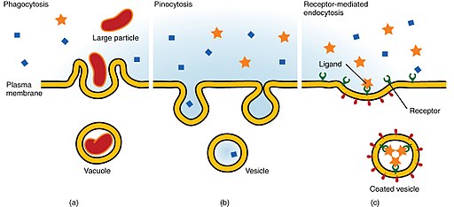
Both phagocytosis and pinocytosis are forms of endocytosis. As such, they involve the transportation of materials from the extracellular matrix through the invagination of the cell membrane. In both pinocytosis and phagocytosis, energy is used for the formation of vesicles.
For this reason, the two are considered forms of active transport. While there are several similarities between pinocytosis and phagocytosis, the two modes of transport are different in a number of ways.
See also: Endocytosis Vs Exocytosis
The following are some of the main differences between the two:
Cell eating vs. cell drinking - One of the main differences between pinocytosis and phagocytosis is with regards to the type of materials they transport into the cell. Whereas phagocytosis involves the ingestion/internalization of solid material (e.g. invading pathogens) and foreign particles, pinocytosis involves the uptake of fluids from the extracellular matrix as well as some solutes.
Because of this difference, pinocytosis is commonly referred to as cell drinking, because extracellular fluid is taken into the cell, while phagocytosis is referred to as cell-eating because solid materials are taken into the cell.
Cell type - While cells are involved in both processes, phagocytosis is especially common in the immune system (in immune cells like granulocytes) while pinocytosis can be observed in different types of cells in the body (endothelial cells, epithelial cells, immune cells, etc.).
Among immune cells, phagocytosis allows for foreign particles, various invading organisms, as well as damaged cells to be ingested and degraded. On the other hand, different types of cells use pinocytosis to take in extracellular fluids along with dissolved solutes into the cell.
For instance, through pinocytosis, cells can take in extracellular fluids as well as proteins required by the cell. Therefore, the two processes are used by different types of cells for different functions.
Pinocytosis is a constant/continuous process - As mentioned, there are various types of pinocytosis that occur in different types of cells. While some of these processes are dependent on receptors and other proteins, pinocytosis has been shown to be a constant/continuous process that allows cells to take in extracellular fluids and various solutes.
This can also be attributed to the fact that for the most part, pinocytosis is nonspecific. For this reason, it continually transports various solutes present in the extracellular fluid along with the inducing molecules/ligands.
Phagocytosis, as well, only occurs when certain material triggers the cell to respond. In the body, for instance, monocytes will only respond in the presence of given activators. For this reason, the process is not continuous.
Vesicle size - In general, smaller vesicles are formed during pinocytosis compared to those formed in phagocytosis. This allows phagocytes to ingest relatively larger particles (cell debris as well as whole invading microorganisms).
Pinocytosis Vs Endocytosis
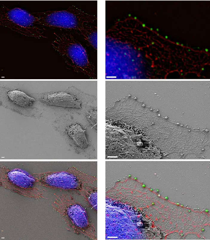
 Zeiss Microscopy:Macrophage endocytosis with correlative superresolution microscopy by https://creativecommons.org/licenses/by-nc-nd/2.0/ no changes made.
Like phagocytosis and Receptor-mediated endocytosis, pinocytosis is also a type of endocytosis. Here, there are several factors that distinguish pinocytosis from the other two types of endocytosis.
Given that differences between pinocytosis and phagocytosis are highlighted in the above section, this section will compare pinocytosis to Receptor-mediated endocytosis:
Type of materials - As mentioned, pinocytosis involves the ingestion/uptake of extracellular fluid and small solutes into the cell. Receptor-mediated endocytosis involves the uptake of specific molecules into the cell. Here, only molecules/material specific to the receptors are transported into the cell through the membrane.
Receptors - As the name suggests, Receptor-mediated endocytosis is dependent on receptors for the uptake of given materials into the cell. Here, specific material/molecules have to bind to the surface receptors in order to be transported.
In pinocytosis, however, receptors do not play the primary role in the transport of materials. Here, pits formed on the membrane will take up fluids and various solutes in the fluid into the cell. For instance, as already mentioned, Clathrin and Caveolae-independent pinocytosis are not dependent on receptors and other coat proteins.
Specificity - Given that receptor-mediated endocytosis is dependent on receptors (on which solutes/molecules bind), the process is selective as only certain substances (those identified by the receptor) can be transported.
As receptors do not play a primary role in pinocytosis, this process is not selective and thus allows for a variety of material to be transported.
Return from Pinocytosis to MicroscopeMaster home
References
Anna L Kiss and Erzsébet Botos. (2009). Endocytosis via caveolae: alternative pathway with distinct cellular compartments to avoid lysosomal degradation?
Kirsten Sandvig, Simona Kavaliauskiene, Tore Skotland. (2018). Clathrin-independent endocytosis: an increasing degree of complexity.
Marta Miaczynska and Harald Stenmark. (2008). Mechanisms and functions of endocytosis.
Links
https://www.bioexplorer.net/pinocytosis.html/#2_Macropinocytosis
https://www.biologyonline.com/dictionary/pinocytosis#Examples_of_Pinocytosis
Find out how to advertise on MicroscopeMaster!


