Endocytosis Vs Exocytosis
** Definitions, Types of Exocytosis
Exocytosis Definition
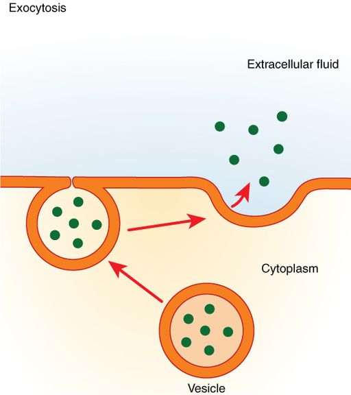
Endocytosis Vs Exocytosis is describing transport. Exocytosis is a type of active transport through which materials are transported and released into the extracellular environment (or transported to the plasma membrane).
Here, materials (waste products, recycled materials, or important substances secreted by the cell, etc.) are first contained within a vesicle. The vesicle (within the cell) then moves to and fuses with the cell membrane where it opens up to release its content.
Compared to some of the substances that can easily diffuse out of the cell through the cell membrane (e.g. oxygen and carbon dioxide etc.), others, like some hormones are too large (macromolecules), and therefore require specialized transportation via transport vesicles.
Different types of cells rely on this form of bulk transport to move material into the extracellular matrix or the surrounding cell membrane.
This is important for a number of functions including:
Cell communication - By producing and releasing signaling molecules, cells can send signals to other cells allowing them to respond. An example of this is the release of neurotransmitters (e.g. dopamine and gamma-Aminobutylic acid etc.) by neurons which are then received by other neurons (postsynaptic neurons)
Recycling - During endocytosis, some receptors may be internalized along with the material of interest into the cell. Through exocytosis, some of these receptors are recycled and transported back to the cell membrane.
Removal of waste material from the cell - To remove some waste material (e.g. lactic acid and some nitrogenous compounds etc.) from the cell, they are contained in vesicles and transported to the extracellular membrane through exocytosis
Other cell secretions - A variety of other products secreted by different cells are transported out of the cell through exocytosis. Some of these secretions include enzymes, some proteins, and antibodies among others
Types of Exocytosis
Though exocytosis generally involves the transportation of material from the cell to the extracellular matrix (or to the plasma membrane), there are several types of exocytosis based on how substances/products are produced.
Constitutive Exocytosis (default pathway)
Constitutive exocytosis is a continuous trafficking pathway through which newly synthesized molecules, especially proteins, are transported to the extracellular matrix. This type of exocytosis occurs in all cells where molecules like proteins have to be transported to the outer surface of the cell (from the Golgi apparatus where they are packaged).
Once materials are synthesized, modified, and transported through the endoplasmic reticulum, they are first packaged in the Golgi apparatus. The membrane of the Golgi apparatus is then used to form new vesicles to contain synthesized materials (e.g. proteins and lipids). The vesicle then pinches off the Golgi body and moves towards the cell membrane in order to release its contents to the outer surface of the cell.
For the most part, the movement of vesicles from the Golgi apparatus to the cell membrane is made possible by microtubules. Here, kinesin or dynein (motor proteins) move along the microtubules in a single direction moving the vesicles in the process.
As well, studies have shown myosin II and myosin V motors to also move vesicles to the cell membrane along the actin cytoskeleton.
Following transportation to the cell membrane, the vesicles go through several key steps that ultimately result in the release of contents into the extracellular matrix. These include tethering and docking where the vesicle links to and attaches to the cell membrane.
Eventually, the vesicles fuse with the cell membrane which opens up the vesicle allowing contents to be released. These steps are regulated by GTPases (enzymes of the Rab family), associated effectors as well as SNARE proteins (large proteins involved in vesicle fusion).
Do animal cells have vesicles?
* A number of proteins play an important role in vesicle formation. During vesicle formation, these proteins (coat proteins) associate with transmembrane proteins consequently pulling the membrane (from Golgi apparatus) into a spherical morphology. Some of the proteins involved in this process include COPII, COPI, and clathrin.
* Some vesicles move from the Golgi apparatus to the endoplasmic reticulum. This allows some materials to be returned back to the endoplasmic reticulum where they belong. A good example of this is the enzyme Protein disulfide-isomerase which is found in the endoplasmic reticulum.
Constitutive exocytosis is an important type of exocytosis that allows cells to constantly transport different types of components to the cell surface to remove them from the cell. For instance, this type of exocytosis plays an important role in continual transport of receptors to the cell membrane. This is especially important in that it ensures that protein receptors are recycled and re-used on the cell surface.
Some of the other material transported through constitutive exocytosis (collectively known as constitutive cargo) include:
· Serum
· Milk proteins
· Proteoglycans
Regulated Exocytosis
The second type of exocytosis is known as regulated exocytosis. Unlike constitutive exocytosis, which is a continual process, regulated exocytosis is dependent on external signals and only occurs when a cell is stimulated/triggered by a signal.
For this reason, this type of exocytosis is mostly common among specialized cells responsible for the secretion of hormones, enzymes, or neurotransmitters among others. Some of the cells characterized by regulated exocytosis include mast cells, neurons, endocrine, and exocrine cells among others.
Because contents of regulated exocytosis are only released when the cell is triggered, vesicles (containing secreted material) are stored in the cell cytoplasm in large numbers until the cell is stimulated.
Regulated Exocytosis in the Islets of Langerhans
Islets of Langerhans are pancreatic cells (beta islet cells) involved in the secretion of insulin (peptide hormone that regulates the breakdown of carbohydrates, proteins, and fats) and glucagon (peptide hormone causes increased concentration of glucose and fatty acids in the bloodstream).
Among other secretory cells, islets of Langerhans are some of the cells in which regulated exocytosis has been well studied. Like many other proteins, insulin precursors (preproinsulin) are produced in ribosomes.
This precursor then undergoes further modification in the endoplasmic reticulum resulting in the production of insulin. Along with another peptide, insulin is packaged in the Golgi apparatus into secretory granules that are released into the cytoplasm.
As mentioned, exocytosis in these cells does not occur until the cell is stimulated. Here, one of the factors that trigger the release of insulin is a rise in blood glucose concentration. Once the cells detect a high concentration of glucose in blood, there are five main steps that take place to release insulin.
These include:
· Trafficking - This is the stage in which the vesicle starts moving towards the membrane along microtubules
· Tethering - The vesicle comes in contact with the membrane
· Docking - Membranes of the cell and the vesicle starts to merge/attach
· Priming - Involves some modification of the cell membrane in response to the external signal
· Fusion - Occurs when the membrane of the cell and that of the vesicle fuse together allowing for vesicular contents to be released. Here, the vesicle may completely fuse with the cell membrane (becoming part of the membrane) or temporarily fuse before separating to return into the cell.
* Some contents of vesicles involved in regulated exocytosis are secreted in the cytosol.
* The type of fusion in which the vesicle does not completely/fully fuse with the cell membrane is known as kiss-and-run fusion.
Lysosome Mediated Exocytosis
Compared to the other types of exocytosis, lysosome mediated exocytosis is the type of exocytosis where the transporting vesicles fuse with the lysosomes. This is an important process through which cells can degrade various material during cellular clearance in order to maintain cell homeostasis.
In addition to cellular clearance, the process has also been associated with a number of other important activities including plasma membrane repair, bone resorption, antigen presentation, as well as contributing to melanocyte functions.
While this type of exocytosis serves a number of important functions, this section will focus on membrane repair to highlight some of the steps involved.
Lysosome Mediated Exocytosis and Membrane Repair
The cell membrane plays an important function in regulating the movement of substances in and out of the cell as well as containing various cell organelles. Therefore, damage to the cell membrane can have significant impacts on a variety of cellular processes.
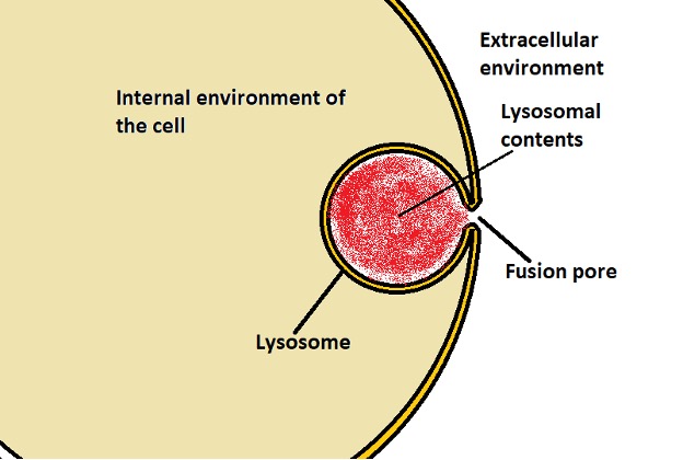
Following damage to the membrane, studies have shown lysosomes (those located near the damaged site) to move and fuse with the cell membrane thereby repairing the damaged part. This is a Ca2+ dependent process that is influenced by an influx of calcium ions following damage to the membrane.
With increased influence of calcium ions through the damaged part, a lysosomal membrane calcium-sensor known as synaptotagmin VII undergoes conformational changes before interacting with SNARE proteins and phospholipids of the cell membrane.
This results in the formation of a fusion pore which not only allows for contents of the lysosome to be released but also for the damaged site to be repaired through sealing.
Endocytosis Vs Exocytosis
Whereas exocytosis involves the transport of material from the cell to the extracellular matrix or to the cell membrane, endocytosis moves material from the extracellular environment into the cell.
This is an important process that serves a number of vital functions including:
Nutrient intake - Cells take up nutrients (e.g. sugars and lipids etc.) through endocytosis. This is important for metabolism and production of ATP energy
Phagocytosis - Immune cells (phagocytes) are able to take ingest invading microbes or other harmful foreign particles through endocytosis. This is known as phagocytosis
Ingestion of old or damaged cells - Immune cells also dispose of dead/damaged cells through endocytosis
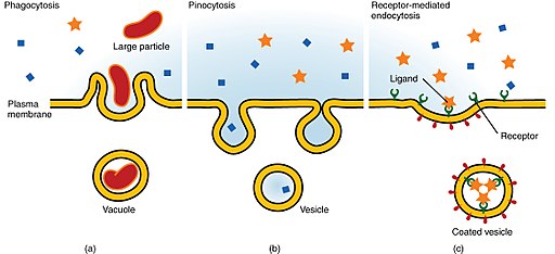
There are three main types of endocytosis which include:
Phagocytosis - Also known as "cell eating" phagocytosis refers to the process through which cells engulf large particles/molecules (generally larger than 0.5um in size). This is therefore an important mechanism through which cells ingest/engulf invading microbes, dead/damaged cells, as well as nutrients, etc.
Pinocytosis - Also known as "cell drinking", pinocytosis is the process through which cells take in small suspended particles from the extracellular matrix. In this case, the cell takes in small suspended/dissolved particles along with some fluid from the extracellular matrix.
Receptor-mediated endocytosis (clathrin-mediated endocytosis) - In this type of endocytosis, different types of molecules and particles like viruses are engulfed and internalized when they come in contact with the cell receptors. Here, clathrin proteins are involved in the formation of membrane vesicles during internalization of molecules/particles.
Endocytosis Vs Exocytosis - Similarities
Active transport - Both exocytosis and endocytosis are active modes of transport. As such, energy is required for the material to be transported across the cell membrane. Here, energy in the form of ATP is especially important because it is involved in vesicle formation during endocytosis as well as the fusion of vesicles to the cell membrane during exocytosis.
Vesicles - Both exocytosis and endocytosis involve the formation of vesicles to transport material. Like the plasma membrane, the vesicle consists of a lipid bilayer with hydrophobic and hydrophilic regions.
The vesicles involved in endocytosis are generally formed from the cell membrane while those involved in exocytosis are formed from the membrane of the Golgi apparatus or the endoplasmic reticulum - In both cases; proteins are also involved in the formation of vesicles.
See also:
Do animal cells have vesicles?
Endocytosis Vs Exocytosis - Differences
Functions - While both exocytosis and endocytosis are involved in the transport of bulk material, they serve different functions. Whereas exocytosis involves the transportation of materials (proteins, lipids, etc.) synthesized in the cell to the extracellular matrix of to the cell membrane, endocytosis involves the movement of material from the extracellular environment (outside the cell) into the cell.
By bringing material/molecules into the cell, endocytosis plays an important role in metabolism, degradation of different types of material as well as activating the cell by introducing signaling molecules etc.
Exocytosis, on the other hand, serves to transport synthesized molecules to the cell membrane, remove waste material, send out signal molecules, and repair the cell membrane among other functions.
Vesicle formation - As mentioned, vesicles involved in exocytosis are formed from the Golgi apparatus and endoplasmic reticulum. Those involved in endocytosis, on the other hand, are formed from the cell membrane.
Types of transport - Some of the main types of endocytosis include phagocytosis, pinocytosis, clathrin-mediated endocytosis, and caveolae-mediated endocytosis. Exocytosis, on the other hand, is divided into calcium ions triggered non-constitutive exocytosis (also known as regulated exocytosis) and non-calcium ions triggered constitutive exocytosis.
Balance of Surface Membrane
Whereas vesicles involved in endocytosis are formed from the cell membrane, those involved in exocytosis are formed from the membrane of Golgi apparatus or that of the endoplasmic reticulum.
Given that endocytosis continuously takes away from the cell membrane, there is a danger of the size and morphology of the cell being significantly affected. However, a balance is maintained by exocytosis where the membranes of vesicles involved in exocytosis are added to the cell membrane through fusion.
Learn about the function of Calcium Signaling
Return from Endocytosis Vs Exocytosis to MicroscopeMaster home
References
Georgeta Basturea. (2019). Endocytosis.
Kuo Liang, Lisi Wei, and Liangyi Chen. (2017). Exocytosis, Endocytosis, and Their Coupling in Excitable Cells.
Marta Miaczynska and Harald Stenmark. (2008). Mechanisms and functions of endocytosis.
Nicholas H. Battey, Nicola C. James, Andrew J. Greenland, and Colin Brownlee. (1999). Exocytosis and Endocytosis.
Links
https://www.sciencedirect.com/topics/medicine-and-dentistry/exocytosis
Find out how to advertise on MicroscopeMaster!


