Why is Cell Adhesion important?
Molecules, Proteins, Assay
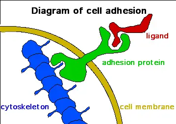
Cell adhesion refers to the process through which a cell forms contact with other cells, substratum in their surroundings, surfaces, as well as the extracellular matrix, etc. Therefore, the term cell adhesion can simply be used to refer to the contact that a cell makes with substances or objects (e.g. glass surface) around them.
This process can be observed in all domains of life including eukarya, archaea, and bacteria. For instance, in eukarya, cell adhesion is evident in living tissue in which bacteria form colonies through cell adhesion.
In nature, cell adhesion is important for various life processes including:
Cell Division and Proliferation
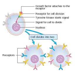
Cell adhesion is an important process for cell division/differentiation given that it heavily influences the polarity and physiological functions of cells, particularly within tissues. Through cell adhesion (cell-to-cell adhesion and cell adhesion to the extracellular matrix etc), cells become part of a microenvironment that consists of other cells and the extracellular matrix. This allows the cell to receive signals that are associated with migration and differentiation etc.
For instance, cell adhesion is required for the leukocyte transmigration during an inflammatory response. This also stimulates proliferation resulting in an increase in the number of cells and becomes even more evident when looking at the impact of antigen-specific immune responses.
While the activation of neutrophils is highly dependent on adhesion with underlying substrate, antigen-specific immune responses stimulate cell division and proliferation to mount a successful attack against the pathogen.
As well, metastatic cancer cells have been shown to have an enhanced ability for cell adhesion. This plays an important role in their migration to new sites where they can establish new tumors. Here, the proliferation of these cells results in the advancement of disease if steps aren't taken to counter and reverse the spread.
Cells that are incapable of adhesion undergo apoptosis within a short period of time as they cannot receive signals from their surroundings. This process has been shown to play an important role in regulating cell division and proliferation.
Infections
Various microorganisms such as certain species of bacteria and Plasmodium species etc are obligate parasites and depend on a host for their survival. For these organisms, cell adhesion is essential for survival given that they heavily depend on their relationship with other organisms.
Using parasitic bacteria as an example, adhesion of these cells to the host cells is the first and most important phase of an infection. For intracellular bacteria (e.g. Salmonella and Brucella species), this creates a bridge that ultimately allows the parasite to invade the cell and enter the cytoplasm where they can obtain their nutrition and multiply.
For other species, however, this allows them to remain in given parts of the body of the host that provides favorable conditions where they can multiply and spread to surrounding tissues. Good examples of this are parasitic bacteria found in the small intestine (e.g. diarrheagenic E. coli).
In the large and small intestine, studies have shown that six (6) stains of diarrheagenic E. coli use different mechanisms to adhere to eukaryotic cells. The 6 pathotypes of E. coli include EPEC, EHEC, ETEC, EAEC, EIEC, and DAEC.
* While cell adhesion is necessary for a successful infection, it's also essential for maintenance of the normal microflora.
Communication (Signaling)
Cell adhesion is very important for communication and signaling. As mentioned, cells rely on signaling in order to respond effectively. Here, then, it's important for cells to remain in contact with each other or with the extracellular matrix in order for signals or information to be transmitted and influence the appropriate responses.
These activities have been observed in tissues where cells are organized in a manner that is appropriate for the tissue function. While cells of the palisade mesophyll are closely packed, cells in the spongy mesophyll are loosely packed in plants.
Regardless, cell adhesion (cell-to-cell and cell to the extracellular matrix, etc) allows for communication and coordination which in turn allows the tissue (consisting of the cells) to perform specific functions that a single cell would otherwise not be able to perform on its own.
In the urinary system, signaling either influences the reabsorption of water or the release of more water in order to conserve or excrete excess water.
Some of the other roles of cell adhesion include:
Cell motility - This is evident through cell adhesion to various surfaces in their surroundings. While some unicellular organisms and even cells of multicellular organisms (e.g. some cells of the immune system) lack such motility structures as flagella, they can move from one region to another through adhesion to substances around them.
Formation of biofilms - Such microorganisms as bacteria and some protists grow on different types of surfaces to form aggregates known as biofilm. This requires different types of cells to stick or adhere to each other. One of the best examples of a biofilm is the dental plaque on teeth's surface.
Growth - Given that cell signaling (made possible by cell adhesion) allows for cell division and proliferation, this permits an organism to grow and increase in size.
Cell Adhesion Proteins and Molecules
Cell adhesion molecules may be described as a family of proteins located on the surface of different types of cells. Although they are all involved in adhesion, as the name suggests, there are different types of molecules that allow their respective cells to respond effectively to external stimuli.
Currently, five main groups/families of adhesion molecules have been identified.
These include:
Integrins
Integrins is a superfamily of adhesion receptors found in various higher (mammals and chicken etc) and lower animals (Drosophila melanogaster, Caenorhabditis elegans, and sponges, etc).
In particular, these adhesion molecules function by binding to ligands (both soluble and cell-surface ligands) and proteins of the extracellular matrix. In higher animals, they have also been shown to play a role in cell-to-cell adhesion in some cases in addition to activating various intracellular signaling pathways.
Although integrins play an important role in cell adhesion, many of them are generally inactive when they are first expressed. For this reason, they cannot bind or participate in signaling when they are newly produced. This is particularly important as it prevents them from performing their respective functions under certain conditions.
The integrin αIIbβ3 found on platelets is inactive in circulating platelets which prevent it from causing thrombosis by binding fibrinogen. Following activation, however, they become active and can perform their respective functions.
Some of the most common integrins include:
- αIIbβ3 located on platelets
- α6β4 located on keratinocytes
- α4β7 found on memory T cells
- αVβ3 found on cells of the endothelium
Integrin Proteins
Like the other adhesion molecules, integrins consist of different types of proteins. Here, the structure consists of alpha and beta subtypes that in turn consist of several regions.
These include:
Alpha subunit - The alpha subunit consists of two important regions that include a thigh and 2 calf domains as well as a domain that consists of about 200 residues.
Whereas the thigh and calf domain support the ligand-binding head, the second region (domain with about 200 residues) is located between the propeller repeats (7 repeats that make up the blade) and is the site that consists of a metal-ion dependent adhesion site involved in ligand binding.
Beta subunit - The beta subunit of integrins consists of four main regions that include the 4 cysteine-rich epidermal growth factor, an I-like domain, the hybrid domain, as well as the plexin-semaphorin-integrin domain.
In addition, it consists of the metal-ion dependent adhesion site which is involved in the binding process.
Selectins
Selectins are a family of adhesion molecules found on various immune cells and the endothelial cells. They include P-selectin found on leukocytes and platelets, E-selectin found on endothelial cells (vascular cells) as well as L-selectin that can be found on other immune cells.
All members of this family are closely related in that they consist of glycoproteins (type I transmembrane glycoproteins). As such, they bind to fucosylated carbohydrates such as P-selectin glycoprotein ligand-1 located on the surface of endothelial cells.
In the body, selectins are involved in a number of processes including homing of stem cells of the bone marrow, inflammation as well as lymphocyte homing.
Cadherins
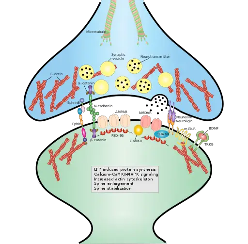
As the name suggests, cadherins are calcium-dependent adhesion molecules. They are some of the most common molecules that form junctions between cells and are therefore involved in cell-to-cell adhesion.
In the process, cadherins play a central role in tissue morphogenesis (tissue development) and homeostasis (internal balance). As mentioned, cell adhesion is important for communication and signaling while communication/signaling is dependent on cell adhesion.
Here, then, communication and signaling influence cell growth, division, and differentiation which in turn regulate tissue development through the formation of distinct cell layers.
In general, the functions of adhesion molecules have been divided into the following categories:
Adhesion tension - Cadherins are directly involved in contact between cells and thus help reduce surface tension at the interface between two cells through adhesion tension. Here, the binding of cadherin over contact results in an increase in adhesion tension which in turn generates a negative tension allowing for the area of contact to expand.
Adhesion signaling - Based on molecular studies, cadherin trans-binding has been shown to result in chemical modifications (e.g. the accumulation of phosphatidylinositol (3,4,5)-trisphosphate). Changes in the chemical composition influence a range of cellular processes such as cytoskeletal rearrangements or uptake of material, etc. Here, then, cadherins are directly involved in signaling.
Mechanical scaffolding/coupling - This is achieved by the anchoring of cadherins to actomyosin cytoskeleton. Here, the adhesion molecules (cadherins) transmit forces to the interconnected cytoskeleton through the cytoskeletal anchor when under a mechanical load.
Although they have similarities, cadherins are divided into four main groups that include:
· Classical cadherins - These include the E, N, and P cadherins (N cadherin is divided into N-cadherin and N-cadherin 2) - These cadherins are characterized by a similar structure.
· Desmosomal cadherins - These are cadherins that form desmosomes. They include desmoglein and desmocollin.
· Protocadherins - Protocadherins are characterized by cadherin motif repeats (five repeats) which distinguish them from the other cadherins.
· Unconventional cadherins - this group consists of different types of cadherins that do not fit into the other groups. Some examples of unconventional cadherins include R-cadherin and VE-cadherin.
Cadherin Proteins
Due to the diversity of cadherins, these adhesion molecules consist of different types of proteins that include:
- CDH1 (Cadherin-1) - Found in E-cadherin
- CDH2 - (Cadherin-2 r neural cadherin) - N-cadherin
- VE-cadherin - Vascular endothelial cadherin
- Desmocollin - A member of the desmosomal cadherins
* Some of the other cadherin proteins include; CDH17, CDH8, CDH3, CDH11, PCDH20, CDH17, and CDH12 etc.
Immunoglobulins
Also known as antibodies, immunoglobulins are glycoproteins typically produced by plasma cells.
It's worth noting that this is a large superfamily that consists of different types of adhesion molecules which include:
Vascular and neural cell adhesion molecules - Vascular adhesion molecules are made up of vascula adhesion protein 1 (VAP-1) and are commonly found in the human endothelial cells. Neural cell adhesion molecule (CD56), on the other hand, consist of glycoproteins and can be found on the surface of skeletal muscle, glia, as well as neurons where they are involved in cell-to-cell adhesion as well as cell recognition.
Intercellular adhesion molecules - Expressed by endothelial and immune cells (some can be found in the brain), intercellular adhesion molecules (ICAM) are made up of intercellular adhesion molecule type 1 proteins and are involved in adhesion as well as the transmigration of leukocytes.
Nectins and nectin-like proteins - Nectins and nectin-like molecules are adhesion molecules that do not require calcium for cellular adhesion. Expressed by different types of cells, these molecules are involved in cell-to-cell adhesion as well as creating adherent junctions.
Mucin-like Molecules
Essentially, mucins are transmembrane glycoproteins that cover some cells and thus cover and affect the functions of various surface molecules. As well, mucin-like molecules such as CD34, GlyCAM-1, and PSGL-1 are found on the surface of some cells and aid in cell adhesion. For instance, CD34 can be found on some endothelial cells where they are involved in binding L-selectin on leukocytes.
Consisting of phosphoglycoprotein, this molecule is involved in the identification of surface antigen which in turn allows for the destruction of associated pathogens. PSGL-1, found on the surface of neutrophils binds to the members of selectin family (E-selectin and P-selectin)
Assay
Cell adhesion assays are used for testing or measuring the cell adhesion capacity of different types of cells. It can be used for a number of functions including measuring the metastatic ability of cancer cells as well as the effects of various treatments based on the ability of cells to adhere.
These assays are important in that they can inform researchers about the state of cells in relation to their functions or given health conditions etc. There are a number of techniques that can be used to measure test adhesion (depending on the cells of interest). In this section, however, we focus on fibronectin cell adhesion assay which is one of the most commonly used assays.
Requirements
- Cells of interest
- 48-well plate
- Phosphate-buffered saline
- Incubator
- Staining solution
- ELISA plate reader
- Deionized water
- Absorbent pad
- Orbital shaker incubator
Procedure
· The pre-coated 48-well plate is first allowed to warm to room temperature
· Once the plate has warmed, it's rinsed in PBS (Phosphate-buffered saline)
· The cells of interest are seeded in the plate and cultured for between 30 and 90 minutes at 37 degrees C
· The culture medium is removed and the cells rinsed 3 to 5 times with PBS
· 200ul of freshly diluted Phosphate-buffered saline is added to each well in the PBS for about 10 minutes (this step is used for fixing)
· After 10 minutes of fixing, this solution is discarded and the cells rinsed with PBS three (3) times
· In each of the well, 200ul of the staining is then added and incubated for about 30 minutes at room temperature (this is done on an orbital shaker for effective staining)
· Once the staining process is completed, the plates are washed using deionized water between 3 and 5 times
· To dry, the plate is inverted onto an absorbent pad
· Lastly, 200ul of the extraction solution is added into each well and the plate incubated for about 5 minutes
* At the end of this protocol, an ELISA plate reader is used to read the results.
Here, learn more about Cell Division, Cell Differentiation, Cell Proliferation , Cellular Metabolism and Pentose Phosphate Pathway
See articles on Cell Culture, Cell Staining and Gram Stain.
What are the Differences between a Plant Cell and an Animal Cell?
Check out information on Cell Biology and Cell Theory.
Return from Why is Cell Adhesion important? to MicroscopeMaster home
References
Alain Carre and Kash L. Mittal. (2011). Surface and Interfacial Aspects of Cell Adhesion.
Alfredo G. Torres Xin Zhou, and James B. Kaper. (2005). Adherence of Diarrheagenic Escherichia coli Strains to Epithelial Cells.
Heidi Harjunpää, et al. (2019). Cell Adhesion Molecules and Their Roles and Regulation in the Immune and Tumor Microenvironment.
Jean-Léon Maître and Carl-Philipp Heisenberg. (2013). Three Functions of Cadherins in Cell Adhesion.
Ley K. (2001) Functions of Selectins. In: Crocker P.R. (eds) Mammalian Carbohydrate Recognition Systems. Results and Problems in Cell Differentiation, vol 33. Springer, Berlin, Heidelberg.
Sceincell Research Laboratories. Cell Adhesion Molecules and Their Roles and Regulation in the Immune and Tumor Microenvironment.
Yoshikazu Takada, Xiaojing Ye & Scott Simon. (2007). The integrins.
Links
https://www.nature.com/scitable/topicpage/cell-adhesion-and-cell-communication-14050486/
Find out how to advertise on MicroscopeMaster!


