What is Pseudomonas Bacteria?
** Characteristics, Gram stain & Infection
Pseudomonas is a genus under the family Pseudomonadaceae. It consists of a large number of species (Gram-negative bacteria species) that exhibit different types of catabolic and metabolic characteristics.
Because of this diversity, members of the genus Pseudomonas have varying relationships with several living organisms including human beings, plants, and different types of animals (cows, dogs, horses, etc). Moreover, Pseudomonas species are also widely distributed in nature and can be found in different ecological niches.
In human beings, animals, and plants, these bacteria can cause infections and diseases as opportunistic pathogens and have therefore received significant attention in medical sciences, agriculture/horticulture, and veterinary medicine.
Some members of the Genus Pseudomonas include:
- Pseudomonas aeruginosa
- Pseudomonas cepacia
- Pseudomonas gladioli
- Pseudomonas facilis
- Pseudomonas pickettii
- Pseudomonas maltophilia
Characteristics
Morphology and Structure
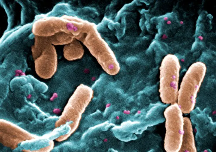
With regards to morphology, Pseudomonas species are rod-shaped and may be straight, slightly curved, or helical. Pseudomonas aeruginosa, which is one of the most common species in this group, for instance, appears to be slightly curved.
Apart from some variation in their general appearance, they also vary in size, ranging from 0.5 to 1.0um in diameter and 1.5 to 5.0um in length.
One of the other morphological characteristics of Pseudomonas species is the presence of one or several flagella. For instance, Pseudomonas aeruginosa has a single unsheathed (lacks an out membranous sheath) located on one polar end of the cell.
For this bacteria, Pseudomonas aeruginosa, the structure serves a number of important functions ranging from swimming motility and adhesion to biofilm formation. The flagellum or flagella in some species, is made up of polymerized flagellin proteins and contents may vary from one species to another.
While some strains of Pseudomonas aeruginosa move by means of a single flagellum, some strains have been shown to possess two or three flagella.
Apart from flagella, Pseudomonas also possesses pili on their surface. Pseudomonas aeruginosa, for instance, have polar type IV pili used for adhesion and twitch motility. In addition, these structures (pili type IV (T4P)) have also been shown to play an important role in pathogenesis and DNA uptake and thus promote bacterial virulence.
Here, it's worth noting that there are four different types of type IV pili. These include IVa, type IVb, and Tad. While they are primarily involved in movement and attachment, some studies suggest that among clinical isolates, pili may have antiphagocytic functions.
Cell structure - Like many other bacteria, Pseudomonas species have a simple cell structure as they lack membrane-bound organelles. However, they have chromosomal DNA and plasmids which have been associated with various advantageous traits in clinical isolates.
Based on various studies, species like Pseudomonas aeruginosa have also been shown to produce extracellular DNA which serves as a cell-to-cell interconnecting matrix component. As compared to chromosomal DNA and plasmids which are intracellular DNA (iDNA), the extracellular DNA (exDNA) can be found outside the cell and is also ubiquitous in nature.
As one of the components that contribute to biofilms, this type of DNA also plays an important role in protecting the bacteria from some cells of the immune system.
Cell envelope - Like Pseudomonas aeruginosa, the majority of Pseudomonas species consist of three main layers covering the cell. These include the inner membrane (also known as the cytoplasmic membrane), the outer membrane as well as the peptidoglycan layer which is located between the inner and outer membrane.
The outer membrane, surrounding the peptidoglycan layer, is made up of several components including protein, lipopolysaccharide (LPS), as well as phospholipids.
While the lipopolysaccharide of pathogenic species like Pseudomonas aeruginosa is toxic, it's generally less toxic when compared to the LPS of various other Gram-negative bacilli. This is one of the reasons a larger dose or inoculum of the bacteria is required to cause a successful infection compared to some of the other Gram-negative pathogens.
* For the majority of Pseudomonas aeruginosa, the lipopolysaccharide consists of several components including hydroxy fatty acids, heptose, core polysaccharides, and 2-keto-3-deoxyoctonic acid.
The peptidoglycan (PG), which is the middle layer (located between the outer and inner membrane), is the main component of the cell wall and consists of Nacetylglucosamine and N-acetylmuramic acid in stem peptide.
Components of the peptidoglycan layer are synthesized within the cell in the cytoplasm before they are exported to the periplasm (also known as the periplasmic space, the periplasm is the region between the inner and outer membranes).
The peptidoglycan layer develops and may be modified or degraded depending on the cell cycle or environmental factors. Lastly, the inner membrane consists of a phospholipid bilayer and is therefore more similar to the cell membrane found in eukaryotic cells.
Here, this membrane is also involved in the movement of molecules in and out of the cell.
Ecology and Distribution
As mentioned, members of the genus Pseudomonas are highly diverse organisms. Because of their relationship with different types of organisms, these bacteria are well spread across the planet wherever these organisms can be found. Moreover, given that the majority of these species are free-living, they can be found in all the natural environments.
While some of the species can be found in aquatic environments, both freshwater and marine, others are found in terrestrial habitats. Although some species can be found in terrestrial environments, they generally prefer moist environments and are therefore likely to be found in moist soil, etc.
* Because of their wide distribution in nature, researchers have suggested that Pseudomonas species are capable of adapting to varying environmental factors.
Some of the factors that have been associated with the diversity of these organisms include:
· Ecological causes - E.g. opportunity and competition
· Genetic causes - E.g. Mutation and genetic recombination
* Apart from soil and water, Pseudomonas species can also be found on the surface of plants and animals as well as fruits etc.
Metabolic Characteristics
Pseudomonas species have been shown to have a robust metabolism for a number of substances including amino acids, organic acids, and aromatic compounds.
While they do not metabolize many sugars, they have a metabolic system in place that allows them to convert glucose to 6-phosphogluconate which is one of the adaptations that allows them to use some sugars for energy.
Here, the diversity of these species allows them to obtain their nutrition in a number of ways.
These include:
Saprophytic nutrition - Some of the species like Pseudomonas putida found in marine and terrestrial environments, exist as saprophytes and therefore obtain nutrition from dead (non-living) organic matter.
Here, these bacteria produce enzymes that are used to process these matters to obtain nutrients. In the process, they also contribute to the decomposition process.
Epiphytic nutrition - Epiphytic bacteria include bacteria that are capable of living and proliferating on the surface of plants like epiphytes. Although these bacteria may cause harm to the plant occasionally, the majority of them do not cause harm and only thrive on nutritional material on the surface of some plants.
Pseudomonas fluorescens is a good example of an epiphytic bacterium that is also being studied as a biocontrol agent.
* In addition to producing antibiotics that protect plants from other bacteria species, some strains of Pseudomonas fluorescens have been shown to produce siderophores and even promote plant growth.
As such, they are also classified as symbiotic bacteria due to their mutual relationship with certain plants.
Parasitic Pseudomonas - While members of the genus Pseudomonas are generally free-living organisms, they are also opportunistic pathogens and can therefore cause diseases/infections on higher organisms.
While Pseudomonas syringiae is an opportunistic pathogen of plants, Pseudomonas aeruginosa is an opportunistic pathogen of both plants and animals. As such, they can invade the host and cause harm as they obtain the nutrition they need to grow and reproduce.
Some of the other characteristics of Pseudomonas species include:
· Aerobic and therefore require free oxygen to reproduce - In the absence of oxygen, they can survive if nitrates are available
· Do not produce spores
· With the exception of a few species, they are mostly oxidase positive
· Are catalase-positive
· Pathogenic species like Pseudomonas aeruginosa produce a capsule-like substance known as alginate, which contributes to the pathogenesis of these organisms - allowing them to evade cells of the immune system of the host
· The majority of species secrete a yellow-green siderophore known as pyoverdine under iron-limiting conditions. However, some of the species may produce extra siderophores such as pyocyanin and thioquinolobactin
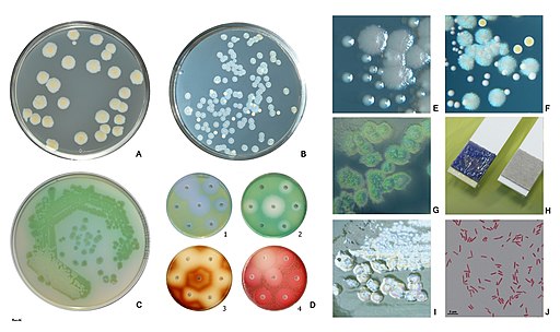
· Depending on the type of species, some of the most common colors of colonies (when grown in standard broth and solid media) are white, yellow, cream, and colorless
· The majority of species are saprophytes
Gram Stain
Gram-stain analysis is one of the most commonly used techniques for the phenotypic characterization of bacteria. Some of the main advantages associated with the method include the fact that it's an easy and quick process that is generally inexpensive.
The goal is to differentiate bacteria based on the capacity of their cell wall with the primary stain (crystal violet) following decolorization (during staining, crystal violet binds to the peptidoglycan of the cell wall).
Given that the bacteria do not usually cause infections in healthy individuals (infections are mild if they occur), samples for analysis may be obtained from infected, hospitalized patients or infected individuals with a compromised immune system.
Samples may include:
· Blood sample - Collected when Pseudomonas cause bacteremia
· Sputum - Collected when Pseudomonas cause an infection in the lungs
· Skin - Skin samples are often used in case of folliculitis (when the bacteria infect the hair follicles)
· Ear discharge - or pus from an eye infection
Following sample collection, a smear is prepared on a glass slide and heat-fixed so that bacterial cells adhere to the slide. Following heat fixing, the slide is flooded with crystal violet (the primary stain) and allowed to stand for about one (1) minute. This allows the stain to bind to the peptidoglycan layer.
The sample is then treated with Gram's iodine which results in the formation of a crystal violet-iodine complex to ensure that the dye is not easily removed. Once the smear is washed gently, it's then treated with a decolorizer, e.g. acetone or ethanol.
The decolorizer serves to dissolve the lipid layer, in the case of Gram-negative bacteria, which allows the primary stain to be removed. In the case of Gram-positive bacteria, the decolorizer dehydrates the thick cell wall which in turn causes the pores to close and prevent the primary stain from leaching out.
It's important to ensure that the decolorization step is prolonged to prevent removal of the stain in both Gram-positive and Gram-negative bacteria.
Following the decolorization step, the smear is again washed before being flooded with the counterstain (secondary stain) like basic fuchsin or safranin. This is particularly important as the secondary stain will stain the Gram-negative bacteria.
Given that the pores of Gram-positive bacteria close during decolorization, they do not take up the secondary stain. However, Gram-negative bacteria easily take up this stain and thus appear pink because they take up the secondary stain.
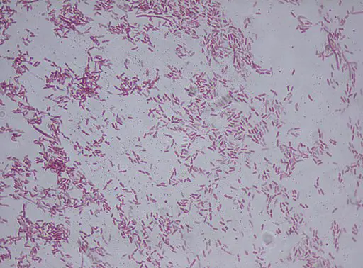
When observed under the microscope, Pseudomonas species will appear pink or reddish (pink rods) in color because they take up the secondary stain. They are classified as Gram-negative bacteria because they are unable to retain the primary stain during decolorization. The majority of these cells occur singly while a few may occur in pairs.
What is Pseudomonas Infection?
Generally, Pseudomonas species are free-living organisms that can be found in water, soil as well as plant surfaces, etc. Although infections in healthy individuals are not common, they tend to be mild when they occur.
As well, infections tend to be more severe among hospitalized patients (nosocomial infection) who have another type of infection as well as individuals with a compromised immune system.
Also known as health-care-associated infections, nosocomial infections occur when the infection exists in a medical setting (and the patient did not have the infection before arriving at the institution).
Some of the groups that are at a high risk of Pseudomonas infections include:
- Diabetes patients
- Patients with severe burns
- Patients with cystic fibrosis
- Patients with HIV/AIDs or an autoimmune disorder
- Patients undergoing chemotherapy
* Being opportunistic pathogens, Pseudomonas species will generally cause disease when the host's immune system has been lowered.
While there are over 140 species of the genus Pseudomonas, about 25 of these have been associated with human beings. Here, some of the most common opportunistic pathogens include Pseudomonas aeruginosa, pseudomonas putida, Pseudomonas putrefaciens, and Pseudomonas fluorescens among others.
Although some of the species were suggested to cause specific diseases in human beings, it's worth noting that these species are no longer considered members of the genus Pseudonomas. Two of these species include Pseudomonas mallei (known today as Burkholderia mallei) and Pseudomonas pseudomallei which is today known as Burkholderia pseudomallei.
* In a clinical setting, Pseudomonas aeruginosa has been shown to be one of the main causes of human infections caused by Pseudomonas. This is largely due to the fact that it's one of the most ubiquitous free-living bacteria.
Given that Pseudomonas species can be found in water, soil, and plants, human beings are highly likely to come in contact with the bacteria.
Some strains of species like Pseudomonas aeruginosa can be found on the skin (particularly around moist regions of the body like the armpit, etc). This makes it easy for the bacteria to cause an infection in the event that the individual's immune system is weakened.
In a hospital setting, the bacteria has been shown to grow well on different types of medical equipment, humidifiers as well as such devices as catheters. If proper care is not taken, it becomes very easy for the bacteria to invade the body of the host and cause disease.
As mentioned, the bacteria may gain entry into the body through wounds (e.g. from surgery), burn wounds, or medical devices, etc.
Some of the most common Pseudomonas infections include:
Folliculitis - Folliculitis is one of the most common skin conditions resulting from the infection of hair follicles. Pseudomonas folliculitis (generally caused by different strains of Pseudomonas aeruginosa) occurs when the bacteria colonize hair follicles following exposure to contaminated water (e.g. in a swimming pool, bathing tubs, and water slides, etc).
However, it may also result from skin contact with contaminated objects like inflatable swimming toys or other bathing objects, etc. This condition is characterized by itchy pustular rashes around the hair follicles
External otitis - External otitis is an infection of the outer ear canal and is also referred to as swimmer's ear. Pseudomonas is the most common cause of the infection and the infection is likely to occur when contaminated water remains in the ear after swimming. In such cases, the water provides a suitable environment for the bacteria to grow and proliferate.
In severe cases (malignant external otitis which has been associated with diabetic patients), the infection is characterized by severe pain in the infected ear, cranial nerve palsies, and may even result in hearing loss. Acute external otitis on the other hand is characterized by itching and tenderness around the ear.
Ecthyma gangrenosum - This is a type of skin lesion that has been observed among patients with a lowered level of neutrophils as well as patients with Pseudomonas aeruginosa (where the bacteria infect the blood). Some of the symptoms of Ecthyma gangrenosum include blisters that quickly transform into pustules and necrotic ulcers.
Pneumonia and respiratory infections - Although Pseudomonas can cause a mild infection in healthy individuals, more severe infections are common among patients who already have other underlying lung conditions (or other respiratory conditions) as well as patients with other health complications.
Among HIV-infected patients, the bacteria have been shown to be one of the main causes of sinutis and pneumonia. On the other hand, the bacteria has been shown to cause ventilator-associated pneumonia as well as bronchitis. Some of the main symptoms of these infections include congestion, severe coughing and chest pains, etc.
Some of the other common Pseudomonas infections include:
Urinary tract infection - E.g. common among individuals using a catheter
Infection of the gastrointestinal tract - For some patients, colonization of the gut by Pseudomonas aeruginosa can result in enteritis. This may be characterized by diarrhea and headaches.
Corneal ulcers - This is an open sore that may result from using contaminated contact lenses etc. It may be characterized by redness of the eyes, excessive tearing, as well as blurry vision in some cases.
Bacteremia - Bacteremia refers to the presence of bacteria in the blood and may result from the use of contaminated IV fluids etc.
See more:
Bacteriology as a field of study
Bacterial Transformation, Conjugation
How do Bacteria cause Disease?
Bacteria - Size, Shape and Arrangement - Eubacteria
Return to Pseudomonas fluorescens
Return to Pseudomonas syringae
Return to Pseudomonas aeruginosa
Return from Pseudomonas to MicroscopeMaster home
References
Andrew J Spiers, Angus Buckling, and Paul B. Rainey. (2000). The cause of Pseudomonas diversity.
Erin M. Anderson et al. (2019). Peptidoglycomics reveals compositional changes in peptidoglycan between biofilm- and planktonic-derived Pseudomonas aeruginosa.
Juan-Luis Ramos. (2004). Pseudomonas: Volume 3 Biosynthesis of Macromolecules and Molecular Metabolism.
Ramakrishnan Sitaraman. (2015). Pseudomonas spp. as models for plant-microbe interactions.
Zulema Udaondo, Juan‐Luis Ramos, Ana Segura, Tino Krell, and Abdelali Daddaoua. (2018).
Regulation of carbohydrate degradation pathways in Pseudomonas involves a versatile set of transcriptional regulators.
Links
Find out how to advertise on MicroscopeMaster!


